Finite-element analysis of the center of resistance of the mandibular dentition
- Affiliations
-
- 1Department of Orthodontics, Graduate School of Clinical Dental Science, The Catholic University of Korea, Seoul, Korea.
- 2Division of Orthodontics, Department of Dentistry, St. Paul's Hospital, College of Medicine, The Catholic University of Korea, Seoul, Korea. dmoss1@hanmail.net
- 3Department of Dentistry, Yonsei University, Seoul, Korea.
- 4Division of Orthodontics, Department of Dentistry, Asan Medical Center, Seoul, Korea.
- 5Department of Orthodontics, Ewha Womans University Mokdong Hospital, Seoul, Korea.
- KMID: 2392193
- DOI: http://doi.org/10.4041/kjod.2017.47.1.21
Abstract
OBJECTIVE
The aim of this study was to investigate the three-dimensional (3D) position of the center of resistance of 4 mandibular anterior teeth, 6 mandibular anterior teeth, and the complete mandibular dentition by using 3D finite-element analysis.
METHODS
Finite-element models included the complete mandibular dentition, periodontal ligament, and alveolar bone. The crowns of teeth in each group were fixed with buccal and lingual arch wires and lingual splint wires to minimize individual tooth movement and to evenly disperse the forces onto the teeth. Each group of teeth was subdivided into 0.5-mm intervals horizontally and vertically, and a force of 200 g was applied on each group. The center of resistance was defined as the point where the applied force induced parallel movement.
RESULTS
The center of resistance of the 4 mandibular anterior teeth group was 13.0 mm apical and 6.0 mm posterior, that of the 6 mandibular anterior teeth group was 13.5 mm apical and 8.5 mm posterior, and that of the complete mandibular dentition group was 13.5 mm apical and 25.0 mm posterior to the incisal edge of the mandibular central incisors.
CONCLUSIONS
Finite-element analysis was useful in determining the 3D position of the center of resistance of the 4 mandibular anterior teeth group, 6 mandibular anterior teeth group, and complete mandibular dentition group.
Figure
-
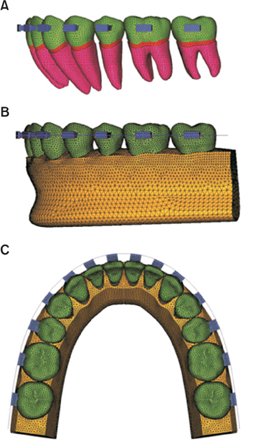
Figure 1 Three-dimensional finite-element mesh applied to the mandibular teeth, periodontal ligaments (PDLs), and alveolar bones. A and B, Lateral views showing the teeth, PDLs, and alveolar bones. C, Occlusal view showing the teeth.
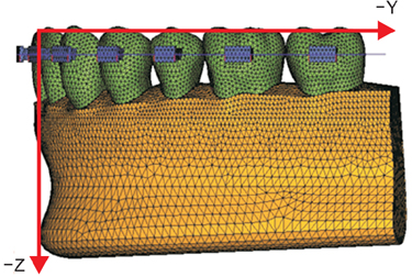
Figure 2 Schematic representation of the coordinate system used in this study.
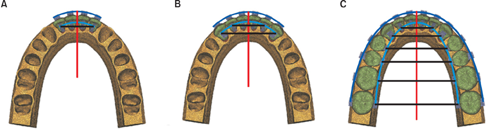
Figure 3 Schematic representation of the finite-element models of the three teeth groups. A, Four mandibular anterior teeth; B, six mandibular anterior teeth; C, the complete mandibular dentition. The blue-colored wires on the buccal and lingual surfaces of the teeth are rigid and have no play with brackets; therefore, the movement of an individual tooth is limited. The black-colored wires cross-linking the left and right teeth are designed to distribute the applied force evenly on the dentition. Vertical force is applied on the red-colored wire.

Figure 4 The contour plot of the mandibular dentition. The original model (white mesh) and deformed model (colored mesh), in which the displacement of the teeth has been magnified 300 times, are superimposed. A, B, and C, Retraction forces were applied at 0, −13.5, and −20 mm, respectively, along the Z-axis. D, E, and F, Intrusion forces were applied at −10, −25, and −40 mm, respectively, along the Y-axis.
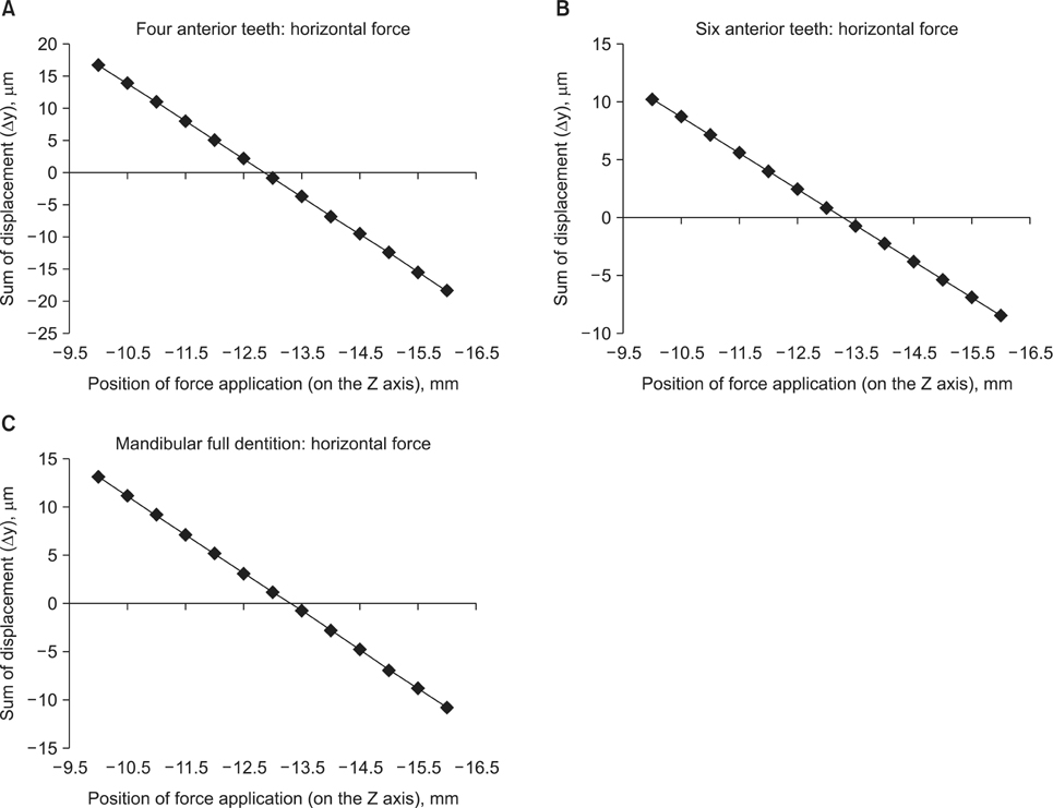
Figure 5 The sum of horizontal displacement (Δy) of the 4 mandibular anterior teeth (A), 6 mandibular anterior teeth (B), and complete mandibular dentition (C) according to variations in the position of force application along the Z-axis.
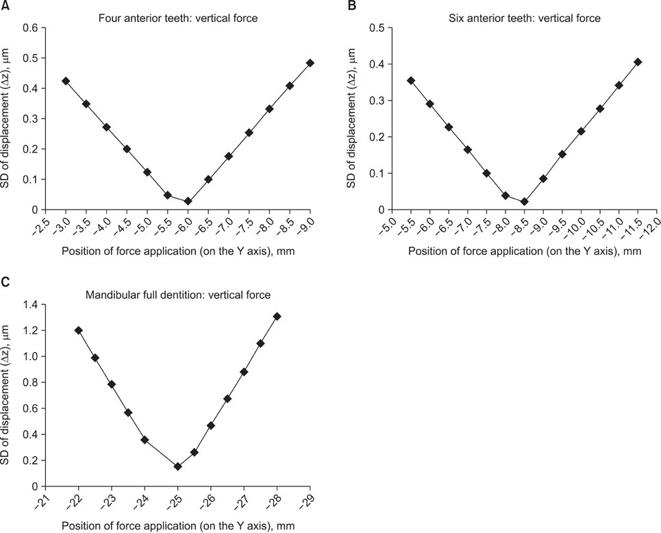
Figure 6 Standard deviation (SD) of vertical displacement (Δz) of the 4 mandibular anterior teeth (A), 6 mandibular anterior teeth (B), and complete mandibular dentition (C) according to variations in the position of force application along the Y-axis.
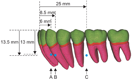
Figure 7 Positions of the centers of resistance. A, The center of resistance of the 4 mandibular anterior teeth; B, the center of resistance of the 6 mandibular anterior teeth; C, the center of resistance of the complete mandibular dentition.
Cited by 2 articles
-
Biomechanical analysis of distalization of mandibular molars by placing a mini-plate: A finite element study
Myungsoon Park, Yonghyun Na, Minbong Park, Janghoon Ahn
Korean J Orthod. 2017;47(5):289-297. doi: 10.4041/kjod.2017.47.5.289.Maxillomandibular arch width differences at estimated centers of resistance: Comparison between normal occlusion and skeletal Class III malocclusion
Yun-Jin Koo, Sung-Hwan Choi, Byeong-Tak Keum, Hyung-Seog Yu, Chung-Ju Hwang, Birte Melsen, Kee-Joon Lee
Korean J Orthod. 2017;47(3):167-175. doi: 10.4041/kjod.2017.47.3.167.
Reference
-
1. Chung KR, Kook YA, Kim SH, Mo SS, Jung JA. Class II malocclusion treated by combining a lingual retractor and a palatal plate. Am J Orthod Dentofacial Orthop. 2008; 133:112–123.
Article2. Lee HK, Chung KR. The vertical location of the center of resistance for maxillary six anterior teeth during retraction using three dimensional finite element analysis. Korean J Orthod. 2001; 31:425–438.3. Reimann S, Keilig L, Jäger A, Bourauel C. Biomechanical finite-element investigation of the position of the centre of resistance of the upper incisors. Eur J Orthod. 2007; 29:219–224.
Article4. Cho SM, Choi SH, Sung SJ, Yu HS, Hwang CJ. The effects of alveolar bone loss and miniscrew position on initial tooth displacement during intrusion of the maxillary anterior teeth: Finite element analysis. Korean J Orthod. 2016; 46:310–322.
Article5. Choi SH, Kim YH, Lee KJ, Hwang CJ. Effect of labiolingual inclination of a maxillary central incisor and surrounding alveolar bone loss on periodontal stress: A finite element analysis. Korean J Orthod. 2016; 46:155–162.
Article6. Choy K, Kim KH, Burstone CJ. Initial changes of centres of rotation of the anterior segment in response to horizontal forces. Eur J Orthod. 2006; 28:471–474.
Article7. Pedersen E, Isidor F, Gjessing P, Andersen K. Location of centres of resistance for maxillary anterior teeth measured on human autopsy material. Eur J Orthod. 1991; 13:452–458.
Article8. Billiet T, de Pauw G, Dermaut L. Location of the centre of resistance of the upper dentition and the nasomaxillary complex. An experimental study. Eur J Orthod. 2001; 23:263–273.
Article9. Park GH, Sohn BW. The center of resistance of the maxillary anterior segment in the horizontal plane during intrusion by using laser reflection technique. Korean J Orthod. 1993; 23:619–631.10. Woo JY, Park YC. Experimental study of the vertical location of the centers of resistance for maxillary anterior teeth during retraction using the laser reflection technique. Korean J Orthod. 1993; 23:375–389.11. Vanden Bulcke MM, Burstone CJ, Sachdeva RC, Dermaut LR. Location of the centers of resistance for anterior teeth during retraction using the laser reflection technique. Am J Orthod Dentofacial Orthop. 1987; 91:375–384.
Article12. Matsui S, Caputo AA, Chaconas SJ, Kiyomura H. Center of resistance of anterior arch segment. Am J Orthod Dentofacial Orthop. 2000; 118:171–178.
Article13. Jeong GM, Sung SJ, Lee KJ, Chun YS, Mo SS. Finite-element investigation of the center of resistance of the maxillary dentition. Korean J Orthod. 2009; 39:83–94.
Article14. Coolidge ED. The thickness of the human periodontal membrane. J Am Dent Assoc. 1937; 24:1260–1270.
Article15. Kronfeld R. Histologic study of the influence of function on the human periodontal membrane. J Am Dent Assoc. 1931; 18:1242–1274.
Article16. Block PL. Restorative margins and periodontal health: a new look at an old perspective. J Prosthet Dent. 1987; 57:683–689.
Article17. Tanne K, Sakuda M, Burstone CJ. Three-dimensional finite element analysis for stress in the periodontal tissue by orthodontic forces. Am J Orthod Dentofacial Orthop. 1987; 92:499–505.
Article18. Jeong HS, Sung SJ, Moon YS, Cho YS, Lim SM. Factors influencing the axes of anterior teeth during SWA en masse sliding retraction with orthodontic mini-implant anchorage: a finite element study. Korean J Orthod. 2006; 36:339–348.19. Chung AJ, Kim US, Lee SH, Kang SS, Choi HI, Jo JH, et al. The pattern of movement and stress distribution during retraction of maxillary incisors using a 3-D finite element method. Korean J Orthod. 2007; 37:98–113.20. Ziegler A, Keilig L, Kawarizadeh A, Jäger A, Bourauel C. Numerical simulation of the biomechanical behaviour of multi-rooted teeth. Eur J Orthod. 2005; 27:333–339.
Article21. Poppe M, Bourauel C, Jäger A. Determination of the elasticity parameters of the human periodontal ligament and the location of the center of resistance of single-rooted teeth a study of autopsy specimens and their conversion into finite element models. J Orofac Orthop. 2002; 63:358–370.
Article22. Andrews LF. Straight wire: the concept and appliance. San Diego, CA: L.A. Wells;1989.23. Germane N, Bentley BE Jr, Isaacson RJ. Three biologic variables modifying faciolingual tooth angulation by straight-wire appliances. Am J Orthod Dentofacial Orthop. 1989; 96:312–319.
Article24. Park CK, Yang WS. A three-dimensional finite element analysis on the location of center of resistance during intrusion of upper anterior teeth. Korean J Orthod. 1997; 27:259–272.25. Bechtold TE, Kim JW, Choi TH, Park YC, Lee KJ. Distalization pattern of the maxillary arch depending on the number of orthodontic miniscrews. Angle Orthod. 2013; 83:266–273.
Article26. Jing Y, Han X, Guo Y, Li J, Bai D. Nonsurgical correction of a Class III malocclusion in an adult by miniscrew-assisted mandibular dentition distalization. Am J Orthod Dentofacial Orthop. 2013; 143:877–887.
Article27. Chung KR, Oh MY, Ko SJ. Corticotomy-assisted orthodontics. J Clin Orthod. 2001; 35:331–339.28. Burstone CJ, Pryputniewicz RJ. Holographic determination of centers of rotation produced by orthodontic forces. Am J Orthod. 1980; 77:396–409.
Article29. Davidovitch M, Rebellato J. Two-couple orthodontic appliance systems utility arches: a two-couple intrusion arch. Semin Orthod. 1995; 1:25–30.
Article30. Choy K, Pae EK, Park Y, Kim KH, Burstone CJ. Effect of root and bone morphology on the stress distribution in the periodontal ligament. Am J Orthod Dentofacial Orthop. 2000; 117:98–105.
Article31. Park HK, Sung EH, Cho YS, Mo SS, Chun YS, Lee KJ. 3-D FEA on the intrusion of mandibular anterior segment using orthodontic miniscrews. Korean J Orthod. 2011; 41:384–398.
Article32. Sung EH, Kim SJ, Chun YS, Park YC, Yu HS, Lee KJ. Distalization pattern of whole maxillary dentition according to force application points. Korean J Orthod. 2015; 45:20–28.
Article
- Full Text Links
-
- Actions
-
Cited
- CITED
-
- Close
- Share
-
- Similar articles
-
- Finite-element investigation of the center of resistance of the maxillary dentition
- The vertical location of the center of resistance for maxillary six anterior teeth during retraction using three dimensional finite element analysis
- Finite-element analysis of the shift in center of resistance of the maxillary dentition in relation to alveolar bone loss
- Three dimensional finite element analysis of mandibular stresses of complete denture occlusion
- A finite element analysis of the center of resistance of a maxillary first molar
