Thoracolithiasis Mimicking a Pleural Plaque in a Patient with a History of Asbestos Exposure: A Case Report
- Affiliations
-
- 1Department of Radiology, Dongguk University Ilsan Hospital, Dongguk University College of Medicine, Goyang, Korea. Jeungkim@dumc.or.kr
- 2Department of Thoracic and Cardiovascular Surgery, Dongguk University Ilsan Hospital, Dongguk University College of Medicine, Goyang, Korea.
- 3Department of Pathology, Dongguk University Ilsan Hospital, Dongguk University College of Medicine, Goyang, Korea.
- KMID: 2392122
- DOI: http://doi.org/10.3348/jksr.2017.77.4.245
Abstract
- A freely mobile calcified or noncalcified nodule in the pleural cavity, known as thoracolithiasis, is quite rare. There are several reports of CT findings of thoracolithiasis, but there is no report of thoracolithiasis mistakenly considered as a pleural plaque in a patient with a history of asbestos exposure. We report a case of a 61-year-old man with a mobile pleural stone thoracoscopically confirmed as thoracolithiasis and which was regarded as a pleural plaque on CT scan in a patient with a history of asbestos exposure.
Figure
-
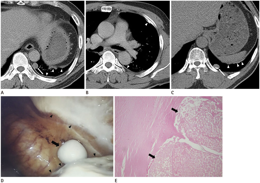
Fig. 1 Thoracolithiasis Mimicking a Pleural Plaque in a 61-year-old man with a History of Asbestos Exposure. A. Contrast-enhanced chest CT scan obtained at the level of the lung base shows a 13-mm sized non-calcified well-defined ovoid nodule (arrow) in the medial portion of the left lower hemithorax. Multiple non-calcified pleural plaques are also noted on the left posterior costal and diaphragmatic pleura (arrowheads). B. Chest CT scan obtained below the level of the carina shows discrete calcified and non-calcified pleural plaques (arrowheads) in the left hemithorax. C. On follow-up evaluation performed eight years later, non-enhanced chest CT shows lateral migration of a pleural nodule (arrow) in the left lower hemithorax and there is newly developed peripheral calcification within the nodule. Pleural plaques are seen again on the left diaphragmatic pleura (arrowheads). D. In the surgical field, a 2 × 1.5 cm sized whitish ovoid hard mass is found floating freely in the pleural cavity (arrow). Whitish pleural plaques are also seen on the parietal pleura (arrowheads). E. Histologic evaluation reveals fatty necrotic tissue (arrows) surrounded by hyalinized fibrous tissue (hematoxylin and eosin; original magnification, × 40).
Reference
-
1. Kinoshita F, Saida Y, Okajima Y, Honda S, Sato T, Hayashibe A, et al. Thoracolithiasis: 11 cases with a calcified intrapleural loose body. J Thorac Imaging. 2010; 25:64–67.2. Kosaka S, Kondo N, Sakaguchi H, Kitano T, Harada T, Nakayama K. Thoracolithiasis. Jpn J Thorac Cardiovasc Surg. 2000; 48:318–321.3. Pineda V, Cáceres J, Andreu J, Vilar J, Domingo ML. Epipericardial fat necrosis: radiologic diagnosis and follow-up. AJR Am J Roentgenol. 2005; 185:1234–1236.4. Iwasaki T, Nakagawa K, Katsura H, Ohse N, Nagano T, Kawahara K. Surgically removed thoracolithiasis: report of two cases. Ann Thorac Cardiovasc Surg. 2006; 12:279–282.5. Kim Y, Shim SS, Chun EM, Won TH, Park S. A pleural loose body mimicking a pleural tumor: a case report. Korean J Radiol. 2015; 16:1163–1165.6. Tanaka D, Niwatsukino H, Fujiyoshi F, Nakajo M. Thoracolithiasis--a mobile calcified nodule in the intrathoracic space: radiographic, CT, and MRI findings. Radiat Med. 2002; 20:131–133.7. Dias AR, Zerbini EJ, Curi N. Pleural stone. A case report. J Thorac Cardiovasc Surg. 1968; 56:120–112.8. Kim JS, Lynch DA. Imaging of nonmalignant occupational lung disease. J Thorac Imaging. 2002; 17:238–260.9. Peacock C, Copley SJ, Hansell DM. Asbestos-related benign pleural disease. Clin Radiol. 2000; 55:422–432.
- Full Text Links
-
- Actions
-
Cited
- CITED
-
- Close
- Share
-
- Similar articles
-
- Asbestosis among Sheetmetal Workers
- Compensation and Diagnosis of Asbestos Related Disease
- The Prevalence of Asbestos Related Pleural Plaque among Residents Living Near Asbestos Mines in Korea
- A Case of Malignant Pleural Mesothelioma Induced by Crocidolite
- CT Findings in People Who Were Environmentally Exposed to Asbestos in Korea
