J Korean Med Sci.
2015 Dec;30(12):1896-1901. 10.3346/jkms.2015.30.12.1896.
CT Findings in People Who Were Environmentally Exposed to Asbestos in Korea
- Affiliations
-
- 1Department of Radiology, Dongguk University Ilsan Hospital, Goyang, Korea. jeungkim@dumc.or.kr
- 2Department of Radiology, Ewha Womans University Mokdong Hospital, Seoul, Korea.
- 3Department of Radiology, Soonchunhyang University Bucheon Hospital, Bucheon, Korea.
- KMID: 2359977
- DOI: http://doi.org/10.3346/jkms.2015.30.12.1896
Abstract
- Asbestos related pleuropulmonary disease has been emerging health problem for recent years. It can cause variable clinical symptoms and radiological abnormalities. However, there has been no report for their characteristics in subjects who were environmentally exposed to asbestos. We reviewed the CT images of 35 people who were environmentally exposed to asbestos in Chungnam province, Korea. The study result showed high incidence of pleural plaque and pulmonary fibrosis on chest CT (94% and 77%, respectively). The common CT findings of lung parenchymal lesions were as follows: centrilobular opacities (94%), subpleural dot-like or branching opacities (80%), interlobular septal thickening (57%), intralobular interstitial thickening (46%), parenchymal bands (43%) and subpleural curvilinear line (29%). There were no significant differences in the prevalence of pulmonary fibrosis and pleural plaques according to sex, age and duration of exposure. In conclusion, pleural plaque and pulmonary fibrosis are common asbestos-related CT finding in the exposed people. Asbestos related lung parenchymal CT findings in the participants with environmental exposure show similar to those observed in the occupational exposure.
Keyword
MeSH Terms
Figure
-
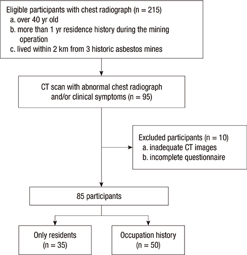
Fig. 1 Flow chart of participant selection.
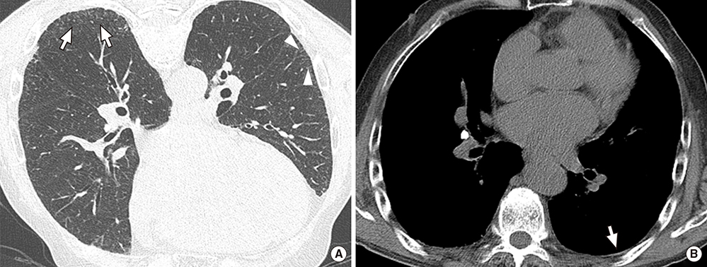
Fig. 2 CT images of a 72-yr-old man with 72 yr residence history. (A) Prone positioned HRCT scan shows subpleural dot-like or branching opacities (arrows), and intralobular interstitial thickening (arrowheads). (B) Noncalcified thin pleural plaque is also noted in left lower posterior portion (arrow).
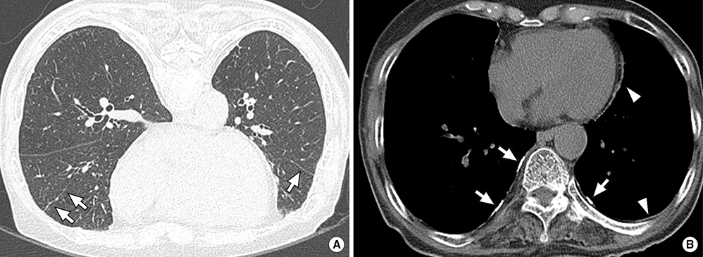
Fig. 3 CT images of a 77-yr-old woman with 69 yr residence history. (A) Prone positioned HRCT scan shows parenchymal band in right mid lobe and left lower lobe (arrows). (B) Multifocal bilateral discontinuous calcified (arrows) and noncalcified (arrowheads) pleural plaques are also noted in mediastinal and bilateral lower posterior areas.
Reference
-
1. Silva CI, Müller NL. Asbestos-related disease. In : Müller NL, Silva CI, editors. Imaging of the chest. Philadelphia: Elsevier Saunders;2008. p. 1140–1166.2. Fraser RS, Colman N, Müller NL, Paré PD. Pulmonary disease caused by inhaled inorganic dust. In : Fraser RS, Colman N, Müller NL, Paré PD, editors. Synopsis of diseases of the chest. 3rd ed. Philadelphia: Elsevier Saunders;2005. p. 714–743.3. Donaldson K, Brown RC, Brown GM. New perspectives on basic mechanisms in lung disease. 5. Respirable industrial fibres: mechanisms of pathogenicity. Thorax. 1993; 48:390–395.4. Lippmann M. Effects of fiber characteristics on lung deposition, retention, and disease. Environ Health Perspect. 1990; 88:311–317.5. Goldberg P, Goldberg M, Marne MJ, Hirsch A, Tredaniel J. Incidence of pleural mesothelioma in New Caledonia: a 10-year survey (1978-1987). Arch Environ Health. 1991; 46:306–309.6. Metintas S, Metintas M, Ucgun I, Oner U. Malignant mesothelioma due to environmental exposure to asbestos: follow-up of a Turkish cohort living in a rural area. Chest. 2002; 122:2224–2229.7. Senyiğit A, Bayram H, Babayiğit C, Topçu F, Nazaroğlu H, Bilici A, Leblebici IH. Malignant pleural mesothelioma caused by environmental exposure to asbestos in the Southeast of Turkey: CT findings in 117 patients. Respiration. 2000; 67:615–622.8. Ökten F, Köksal D, Önal M, Özcan A, Şimşek C, Ertürk H. Computed tomography findings in 66 patients with malignant pleural mesothelioma due to environmental exposure to asbestos. Clin Imaging. 2006; 30:177–180.9. Asbestos, asbestosis, and cancer: the Helsinki criteria for diagnosis and attribution. Scand J Work Environ Health. 1997; 23:311–316.10. Song SH, Hwang JH, Hwang BG, Kim HW. Occurrence types and mineralogical characteristics of asbestos for the Kwangcheon area, Chungnam. J Korean Soc Occup Environ Hyg. 2008; 18:271–281.11. Ahn YS, Kim HR. Asbestosis epidemics caused by non-occupational neighborhood exposure. J Korean Med Assoc. 2009; 52:472–481.12. Kang DM, Gu DC, Kim KH. Asbestos-related diseases among asbestos textile factory workers and residents around the factory. J Korean Med Assoc. 2009; 52:482–488.13. Akira M, Yamamoto S, Yokoyama K, Kita N, Morinaga K, Higashihara T, Kozuka T. Asbestosis: high-resolution CT-pathologic correlation. Radiology. 1990; 176:389–394.14. Akira M, Yamamoto S, Inoue Y, Sakatani M. High-resolution CT of asbestosis and idiopathic pulmonary fibrosis. AJR Am J Roentgenol. 2003; 181:163–169.15. Aberle DR, Gamsu G, Ray CS, Feuerstein IM. Asbestos-related pleural and parenchymal fibrosis: detection with high-resolution CT. Radiology. 1988; 166:729–734.16. Aberle DR, Gamsu G, Ray CS. High-resolution CT of benign asbestos-related diseases: clinical and radiographic correlation. AJR Am J Roentgenol. 1988; 151:883–891.17. Friedman AC, Fiel SB, Fisher MS, Radecki PD, Lev-Toaff AS, Caroline DF. Asbestos-related pleural disease and asbestosis: a comparison of CT and chest radiography. AJR Am J Roentgenol. 1988; 150:269–275.18. Staples CA, Gamsu G, Ray CS, Webb WR. High resolution computed tomography and lung function in asbestos-exposed workers with normal chest radiographs. Am Rev Respir Dis. 1989; 139:1502–1508.19. Akira M, Yokoyama K, Yamamoto S, Higashihara T, Morinaga K, Kita N, Morimoto S, Ikezoe J, Kozuka T. Early asbestosis: evaluation with high-resolution CT. Radiology. 1991; 178:409–416.20. Oksa P, Suoranta H, Koskinen H, Zitting A, Nordman H. High-resolution computed tomography in the early detection of asbestosis. Int Arch Occup Environ Health. 1994; 65:299–304.21. Lynch DA. CT for asbestosis: value and limitations. AJR Am J Roentgenol. 1995; 164:69–71.22. Harkin TJ, McGuinness G, Goldring R, Cohen H, Parker JE, Crane M, Naidich DP, Rom WN. Differentiation of the ILO boundary chest roentgenograph (0/1 to 1/0) in asbestosis by high-resolution computed tomography scan, alveolitis, and respiratory impairment. J Occup Environ Med. 1996; 38:46–52.23. Soulat JM, Lauque D, Esquirol Y, Déprés M, Giron J, Claudel R, Carles P. High-resolution computed tomography abnormalities in ex-insulators annually exposed to asbestos dust. Am J Ind Med. 1999; 36:593–601.24. Weiss W. Cigarette smoke, asbestos, and small irregular opacities. Am Rev Respir Dis. 1984; 130:293–301.25. Blanc PD, Golden JA, Gamsu G, Aberle DR, Gold WM. Asbestos exposure-cigarette smoking interactions among shipyard workers. JAMA. 1988; 259:370–373.26. Barnhart S, Thornquist M, Omenn GS, Goodman G, Feigl P, Rosenstock L. The degree of roentgenographic parenchymal opacities attributable to smoking among asbestos-exposed subjects. Am Rev Respir Dis. 1990; 141:1102–1106.27. Kim I, Kim EA, Kim JY. Compensation for occupational cancer. J Korean Med Sci. 2014; 29:S40–S46.28. American Thoracic Society. Diagnosis and initial management of nonmalignant diseases related to asbestos. Am J Respir Crit Care Med. 2004; 170:691–715.29. Anderson HA, Lilis R, Daum SM, Fischbein AS, Selikoff IJ. Household-contact asbestos neoplastic risk. Ann N Y Acad Sci. 1976; 271:311–323.
- Full Text Links
-
- Actions
-
Cited
- CITED
-
- Close
- Share
-
- Similar articles
-
- Occupational and Environmental Asbestos Exposure in Korea
- Asbestos-related Diseases among Asbestos Textile Factory Workers and Residents Around the Factory
- Types and Health Hazards of Fibrous Materials Used as Asbestos Substitutes
- Compensation and Diagnosis of Asbestos Related Disease
- Systematic Review of the Effects of Asbestos Exposure on the Risk of Cancer between Children and Adults
