J Periodontal Implant Sci.
2017 Aug;47(4):231-239. 10.5051/jpis.2017.47.4.231.
Marginal bone level changes in association with different vertical implant positions: a 3-year retrospective study
- Affiliations
-
- 1Department of Periodontology, Wonkwang University Daejeon Dental Hospital, Wonkwang University College of Dentistry, Daejeon, Korea. seongnyum@wonkwang.ac.kr
- KMID: 2388110
- DOI: http://doi.org/10.5051/jpis.2017.47.4.231
Abstract
- PURPOSE
To retrospectively evaluate the relationship between the vertical position of the implant-abutment interface and marginal bone loss over 3 years using radiological analysis.
METHODS
In total, 286 implant surfaces of 143 implants from 61 patients were analyzed. Panoramic radiographic images were taken immediately after implant installation and at 6, 12, and 36 months after loading. The implants were classified into 3 groups based on the vertical position of the implant-abutment interface: group A (above bone level), group B (at bone level), and group C (below bone level). The radiographs were analyzed by a single examiner.
RESULTS
Changes in marginal bone levels of 0.99±1.45, 1.13±0.91, and 1.76±0.78 mm were observed at 36 months after loading in groups A, B, and C, respectively, and bone loss was significantly greater in group C than in groups A and B.
CONCLUSIONS
The vertical position of the implant-abutment interface may affect marginal bone level change. Marginal bone loss was significantly greater in cases where the implant-abutment interface was positioned below the marginal bone. Further long-term study is required to validate our results.
MeSH Terms
Figure
-
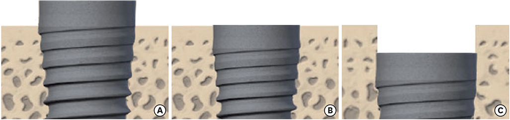
Figure 1 Experimental group classification. (A) Group A; (B) group B; and (C) group C. Group A: implant-abutment interface positioned above the marginal bone, Group B: implant-abutment interface positioned at the marginal bone level, Group C: implant-abutment interface positioned below the marginal bone.
Reference
-
1. Higuchi KW, Folmer T, Kultje C. Implant survival rates in partially edentulous patients: a 3-year prospective multicenter study. J Oral Maxillofac Surg. 1995; 53:264–268.2. Jung RE, Pjetursson BE, Glauser R, Zembic A, Zwahlen M, Lang NP. A systematic review of the 5-year survival and complication rates of implant-supported single crowns. Clin Oral Implants Res. 2008; 19:119–130.
Article3. Pjetursson BE, Asgeirsson AG, Zwahlen M, Sailer I. Improvements in implant dentistry over the last decade: comparison of survival and complication rates in older and newer publications. Int J Oral Maxillofac Implants. 2014; 29:Suppl. 308–324.
Article4. Adell R, Lekholm U, Rockler B, Brånemark PI. A 15-year study of osseointegrated implants in the treatment of the edentulous jaw. Int J Oral Surg. 1981; 10:387–416.
Article5. Albrektsson T, Zarb G, Worthington P, Eriksson AR. The long-term efficacy of currently used dental implants: a review and proposed criteria of success. Int J Oral Maxillofac Implants. 1986; 1:11–25.6. Smith DE, Zarb GA. Criteria for success of osseointegrated endosseous implants. J Prosthet Dent. 1989; 62:567–572.
Article7. Cox JF, Zarb GA. The longitudinal clinical efficacy of osseointegrated dental implants: a 3-year report. Int J Oral Maxillofac Implants. 1987; 2:91–100.8. Oh TJ, Yoon J, Misch CE, Wang HL. The causes of early implant bone loss: myth or science? J Periodontol. 2002; 73:322–333.
Article9. Quirynen M, van Steenberghe D. Bacterial colonization of the internal part of two-stage implants. An in vivo study. Clin Oral Implants Res. 1993; 4:158–161.
Article10. Ericsson I, Persson LG, Berglundh T, Marinello CP, Lindhe J, Klinge B. Different types of inflammatory reactions in peri-implant soft tissues. J Clin Periodontol. 1995; 22:255–261.
Article11. Abrahamsson I, Berglundh T, Lindhe J. Soft tissue response to plaque formation at different implant systems. A comparative study in the dog. Clin Oral Implants Res. 1998; 9:73–79.
Article12. Lindhe J, Hamp SE, Löe H. Plaque induced periodontal disease in beagle dogs. A 4-year clinical, roentgenographical and histometrical study. J Periodontal Res. 1975; 10:243–255.13. Lindhe J, Ericsson I. Effect of ligature placement and dental plaque on periodontal tissue breakdown in the dog. J Periodontol. 1978; 49:343–350.
Article14. Broggini N, McManus LM, Hermann JS, Medina RU, Oates TW, Schenk RK, et al. Persistent acute inflammation at the implant-abutment interface. J Dent Res. 2003; 82:232–237.
Article15. Schwarz F, Hegewald A, Becker J. Impact of implant-abutment connection and positioning of the machined collar/microgap on crestal bone level changes: a systematic review. Clin Oral Implants Res. 2014; 25:417–425.
Article16. Yi JM, Lee JK, Um HS, Chang BS, Lee MK. Marginal bony changes in relation to different vertical positions of dental implants. J Periodontal Implant Sci. 2010; 40:244–248.
Article17. Hermann JS, Schoolfield JD, Schenk RK, Buser D, Cochran DL. Influence of the size of the microgap on crestal bone changes around titanium implants. A histometric evaluation of unloaded non-submerged implants in the canine mandible. J Periodontol. 2001; 72:1372–1383.
Article18. Veis A, Parissis N, Tsirlis A, Papadeli C, Marinis G, Zogakis A. Evaluation of peri-implant marginal bone loss using modified abutment connections at various crestal level placements. Int J Periodontics Restorative Dent. 2010; 30:609–617.19. van Eekeren P, Tahmaseb A, Wismeijer D. Crestal bone changes in macrogeometrically similar implants with the implant-abutment connection at the crestal bone level or 2.5 mm above: a prospective randomized clinical trial. Clin Oral Implants Res. 2016; 27:1479–1484.
Article20. Koo KT, Lee EJ, Kim JY, Seol YJ, Han JS, Kim TI, et al. The effect of internal versus external abutment connection modes on crestal bone changes around dental implants: a radiographic analysis. J Periodontol. 2012; 83:1104–1109.
Article21. Weng D, Nagata MJ, Bosco AF, de Melo LG. Influence of microgap location and configuration on radiographic bone loss around submerged implants: an experimental study in dogs. Int J Oral Maxillofac Implants. 2011; 26:941–946.22. Lazzara RJ, Porter SS. Platform switching: a new concept in implant dentistry for controlling postrestorative crestal bone levels. Int J Periodontics Restorative Dent. 2006; 26:9–17.23. Schopper C, Moser D, Goriwoda W, Ziya-Ghazvini F, Spassova E, Lagogiannis G, et al. The effect of three different calcium phosphate implant coatings on bone deposition and coating resorption: a long-term histological study in sheep. Clin Oral Implants Res. 2005; 16:357–368.
Article24. Xuereb M, Camilleri J, Attard NJ. Systematic review of current dental implant coating materials and novel coating techniques. Int J Prosthodont. 2015; 28:51–59.
Article25. Oshida Y, Tuna EB, Aktören O, Gençay K. Dental implant systems. Int J Mol Sci. 2010; 11:1580–1678.
Article
- Full Text Links
-
- Actions
-
Cited
- CITED
-
- Close
- Share
-
- Similar articles
-
- Marginal bony changes in relation to different vertical positions of dental implants
- A Two-year Retrospective Study on the Clinical Success of the Korean Implant Systems
- Influence of crown-to-implant ratio on periimplant marginal bone loss in the posterior region: a five-year retrospective study
- Radiographic evaluation of the proximal bone level between two implants: A 3-year comparative study between Branemark and ITI implants in the mandibular posterior region
- Effect of the vertical implant position relative to the adjacent cementoenamel junction on peri-implantation bone loss
