Investig Clin Urol.
2017 Sep;58(5):339-345. 10.4111/icu.2017.58.5.339.
Radiological noninvasive assessment of ureteral stone impaction into the ureteric wall: A critical evaluation with objective radiological parameters
- Affiliations
-
- 1Dr. Lütfi Kirdar Training and Research Hospital Radiology Clinic, Istanbul, Turkey. ozlemelibol@ymail.com
- KMID: 2388050
- DOI: http://doi.org/10.4111/icu.2017.58.5.339
Abstract
- PURPOSE
To determine the predictive value of certain radiological parameters for an objective asssessment of the presence of ureteral stone impaction.
MATERIALS AND METHODS
Seventy-nine patients with a single proximal ureteral stones were retrieved from the departmental database. Both clinical and particularly radiological data of all cases were well evaluated on this aspect. In addition to the time period between the first colic attack and definitive management; diameter of proximal ureter and renal pelvis, longitudinal and transverse stone size, Hounsfied unit (HU) of the stone and lastly ureteral wall thickness at the impacted stone site were all carefully evaluated and noted.
RESULTS
Patients had a single proximal ureteral stone. While mean age of the cases was ranged 20 to 78 years; mean stone size was 15.62±4.26 mm. Evaluation of our data demonstrated that although there was a statistically significant correlation between ureteral wall thickness and patients age, transverse diameter of the stone, ureteral diameter just proximal to the stone, renal pelvic diameter and the duration of renal colic attacks; no correlation could be demonstrated between patients sex and the HU of the stone.
CONCLUSIONS
Prediction of the presence and degree of proximal ureteral stone impaction is a challenging issue and our data indicated a highly significant correlation between ureteral wall thickness and the some certain radiological as well as clinical parameters evaluated which will give an objective information about the presence of impaction which may in turn be helpful in the follow-up and also management plans of such calculi.
Keyword
Figure
-
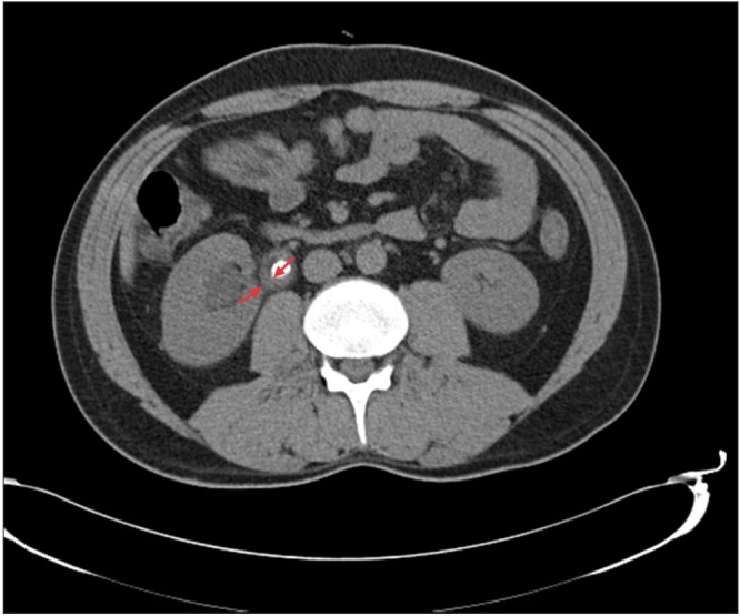
Fig. 1 Measurement of ureteral wall thickness from an axial computed tomographic image (arrows).
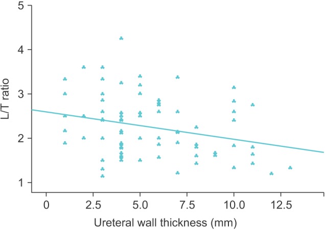
Fig. 2 Evaluation of the relationship between ureteral wall thickness and longitudinal/transverse (L/T) size of the stone ratio.
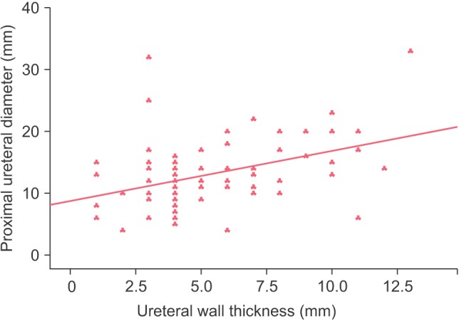
Fig. 3 Evaluation of the relationship between ureteral wall thickness and proximal ureteral diameter.
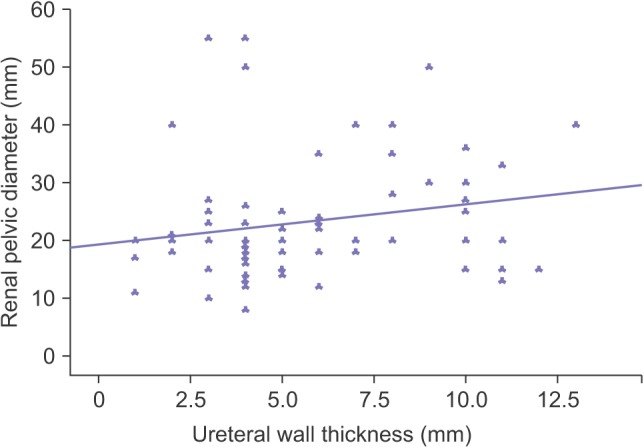
Fig. 4 Evaluation of the relationship between ureteral wall thickness and renal pelvic diameter.
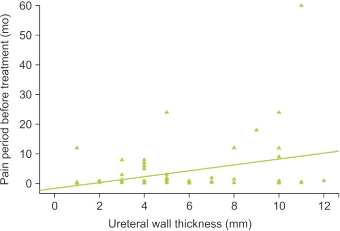
Fig. 5 Evaluation of the relationship between ureteral wall thickness and the pain period before treatment.
Reference
-
1. Mugiya S, Ito T, Maruyama S, Hadano S, Nagae H. Endoscopic features of impacted ureteral stones. J Urol. 2004; 171:89–91. PMID: 14665851.
Article2. Deliveliotis C, Chrisofos M, Albanis S, Serafetinides E, Varkarakis J, Protogerou V. Management and follow-up of impacted ureteral stones. Urol Int. 2003; 70:269–272. PMID: 12740489.
Article3. Morgentaler A, Bridge SS, Dretler SP. Management of the impacted ureteral calculus. J Urol. 1990; 143:263–266. PMID: 1967657.
Article4. Seitz C, Tanovic E, Kikic Z, Fajkovic H. Impact of stone size, location, composition, impaction, and hydronephrosis on the efficacy of holmium:YAG-laser ureterolithotripsy. Eur Urol. 2007; 52:1751–1757. PMID: 17459573.
Article5. Wolf JS Jr. Treatment selection and outcomes: ureteral calculi. Urol Clin North Am. 2007; 34:421–430. PMID: 17678991.
Article6. Takahashi N, Kawashima A, Ernst RD, Boridy IC, Goldman SM, Benson GS, et al. Ureterolithiasis: can clinical outcome be predicted with unenhanced helical CT. Radiology. 1998; 208:97–102. PMID: 9646798.
Article7. Fielding JR, Silverman SG, Samuel S, Zou KH, Loughlin KR. Unenhanced helical CT of ureteral stones: a replacement for excretory urography in planning treatment. AJR Am J Roentgenol. 1998; 171:1051–1053. PMID: 9762995.
Article8. Fowler JC, Cutress ML, Abubacker Z, Saleemi MA, Alam A, Shekhdar J, et al. Clinical evaluation of ultra-low dose contrast-enhanced CT in patients presenting with acute ureteric colic. J Clin Urol. 2011; 4:56–63.
Article9. Varanelli MJ, Coll DM, Levine JA, Rosenfield AT, Smith RC. Relationship between duration of pain and secondary signs of obstruction of the urinary tract on unenhanced helical CT. AJR Am J Roentgenol. 2001; 177:325–330. PMID: 11461855.
Article10. Sarica K, Kafkasli A, Yazici Ö, Çetinel AC, Demirkol MK, Tuncer M, et al. Ureteral wall thickness at the impacted ureteral stone site: a critical predictor for success rates after SWL. Urolithiasis. 2015; 43:83–88. PMID: 25417717.
Article11. Morgentaler A, Bridge SS, Dretler SP. Management of the impacted ureteral calculus. J Urol. 1990; 143:263–266. PMID: 1967657.
Article12. Srivastava A, Ahlawat R, Kumar A, Kapoor R, Bhandari M. Management of impacted upper ureteric calculi: results of lithotripsy and percutaneous litholapaxy. Br J Urol. 1992; 70:252–257. PMID: 1422683.
Article13. Elganainy E, Hameed DA, Elgammal M, Abd-Elsayed AA, Shalaby M. Experience with impacted upper ureteral stones; should we abandon using semirigid ureteroscopes and pneumatic lithoclast. Int Arch Med. 2009; 2:13. PMID: 19409107.
Article14. Juan YS, Shen JT, Li CC, Wang CJ, Chuang SM, Huang CH, et al. Comparison of percutaneous nephrolithotomy and ureteroscopic lithotripsy in the management of impacted, large, proximal ureteral stones. Kaohsiung J Med Sci. 2008; 24:204–209. PMID: 18424357.
Article15. Roberts WW, Cadeddu JA, Micali S, Kavoussi LR, Moore RG. Ureteral stricture formation after removal of impacted calculi. J Urol. 1998; 159:723–726. PMID: 9474134.
Article
- Full Text Links
-
- Actions
-
Cited
- CITED
-
- Close
- Share
-
- Similar articles
-
- Intravenous pyelographic analysis of ureteral calculi
- Ureteral stricture formation after ureteroscope treatment of impacted calculi: A prospective study
- Unilateral ureteric stone associated with gross hydronephrosis and kidney shrinkage: a cadaveric report
- The Comparison of Efficacy of Ureteroscopic Removal and Shockwave Lithotripsy in Lower Ureteral Stones
- Clinical Observation on Cystoscpic Removal of Lower Third Ureteral Stone Using Domina Stone Dislodger
