Ann Clin Neurophysiol.
2017 Jul;19(2):118-124. 10.14253/acn.2017.19.2.118.
Electroencephalography for the diagnosis of brain death
- Affiliations
-
- 1Department of Neurology, Kangwon National University School of Medicine, Chuncheon, Korea.
- 2Department of Neurology, Gangnam Severance Hospital, Yonsei University College of Medicine, Seoul, Korea.
- 3Department of Neurology, Chungnam National University Hospital, Daejeon, Korea.
- 4Department of Neurology, Hanyang University Seoul Hospital, Seoul, Korea.
- 5Department of Neurology, Yonsei University College of Medicine, Seoul, Korea.
- KMID: 2387232
- DOI: http://doi.org/10.14253/acn.2017.19.2.118
Abstract
- Electroencephalography (EEG) is frequently used to assist the diagnosis of brain death. However, to date there have been no guidelines in terms of EEG criteria for determining brain death in Korea, despite EEG being mandatory. The purpose of this review is to provide an update on the evidence and controversies with regarding to the utilization of EEG for determining brain death and to serve as a cornerstone for the development of future guidelines. To determine brain death, electrocerebral inactivity (ECI) should be demonstrated on EEG at a sensitivity of 2 µV/mm using double-distance electrodes spaced 10 centimeters or more apart from each other for at least 30 minutes, with intense somatosensory or audiovisual stimuli. ECI should be also verified by checking the integrity of the system. Additional monitoring is needed if extracerebral potentials cannot be eliminated. Interpreting EEG at high sensitivities, which is required for the diagnosis of brain death, can pose a diagnostic challenge. Furthermore, EEG is affected by physiologic variables and drugs. However, no consensus exists as to the minimal requirements for blood pressure, oxygen saturation, and body temperature during the EEG recording itself, the minimal time for observation after the brain injury or rewarming from hypothermia, and how to determine brain death when the findings of ECI is equivocal. Therefore, there is a strong need to establish detailed guidelines for performing EEG to determine brain death.
MeSH Terms
Figure
-
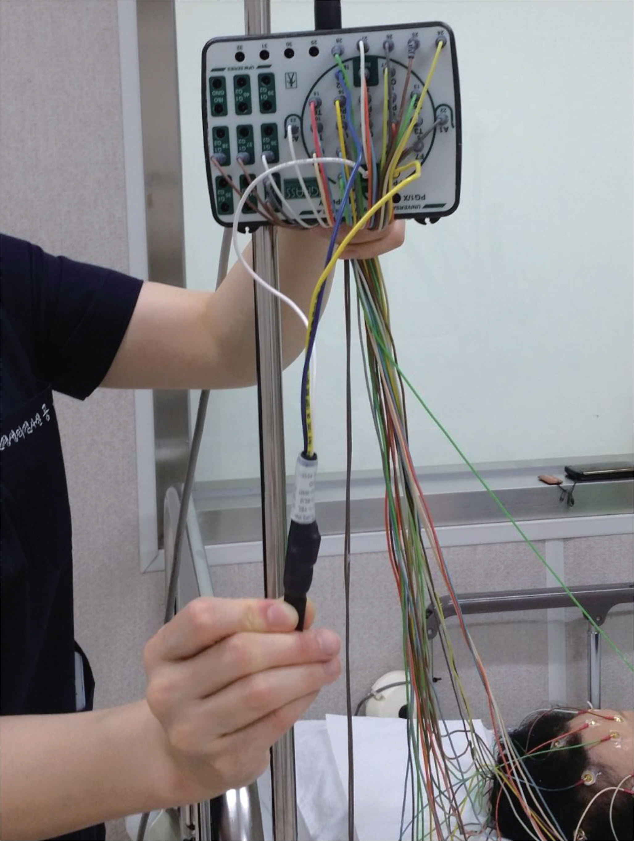
Fig. 1. An example of a dummy patient with a three-lead device (Grass Technologies, West Warwick, RI, USA). This can be used to measure machine noise and external interference which enters to the recording system.

Fig. 2. EEG of a 70-year-old comatose male recorded 3 days after cardiopulmonary arrest. (A) The EEG appeared flat at sensitivity 7 μV/mm without any reactivity to painful stimuli. (B) Low-voltage, mixed-frequency activity was seen at sensitivity 2 μV/mm in the same epoch, which could not be eliminated. The patient never regained consciousness, spontaneous respiration, or brainstem reflexes after the cardiopulmonary arrest. A repeat EEG performed 12 days after the arrest showed the same pattern. EEG, electroencephalography.
Reference
-
1.Practice parameters for determining brain death in adults (sum-mary statement). The Quality Standards Subcommittee of the American Academy of Neurology. Neurology. 1995. 45:1012–1014.2.Wijdicks EF. Brain death worldwide: accepted fact but no global consensus in diagnostic criteria. Neurology. 2002. 58:20–25.
Article3.Wahlster S., Wijdicks EF., Patel PV., Greer DM., Hemphill JC 3rd., Carone M, et al. Brain death declaration: practices and perceptions worldwide. Neurology. 2015. 84:1870–1879.
Article4.Kramer AH. Ancillary testing in brain death. Semin Neurol. 2015. 35:125–138.
Article5.A definition of irreversible coma. Report of the Ad Hoc Committee of the Harvard Medical School to examine the definition of brain death. JAMA. 1968. 205:337–340.6.Nam SO. Brain death and organ transplantation. Korean J Pediatr. 2009. 52:856–861.
Article7.Korean Ministry of Gevernment Legislation Pages©. Enforcement decree of the organ transplant act [Internet]. Sejong, Korea: Korean Ministry of Gevernment Legislation;c2016. [accessed 2016 Oct 28]. Available from. http://www.law.go.kr.8.Korea Organ Donation Agency Pages©. KODA Annual Report 2015 [Internet]. Seoul, Korea: Korea Organ Donation Agency;c2015. [accessed 2016 Oct 28]. Available from. http://fliphtml5.com/down-load-pdf-file/590915.9.American Clinical Neurophysiology Society. Guideline 3: Minimum technical standards for EEG recording in suspected cerebral death. J Clin Neurophysiol. 2006. 23:97–104.10.Stecker MM., Sabau D., Sullivan L., Das RR., Selioutski O., Drislane FW, et al. American Clinical Neurophysiology Society Guideline 6: Minimum technical standards for EEG recording in suspected cerebral death. J Clin Neurophysiol. 2016. 33:324–327.
Article11.A glossary of terms most commonly used by clinical electro-encephalographers. Electroencephalogr Clin Neurophysiol. 1974. 37:538–548.12.André-Obadia N., Lamblin MD., Sauleau P. French recommendations on electroencephalography. Neurophysiol Clin. 2015. 45:1–17.
Article13.Webb AC., Samuels OB. Reversible brain death after cardiopulmonary arrest and induced hypothermia. Crit Care Med. 2011. 39:1538–1542.
Article14.Young GB., Lee D. A critique of ancillary tests for brain death. Neur-ocrit Care. 2004. 1:499–508.
Article15.Brierley JB., Graham DI., Adams JH., Simpsom JA. Neocortical death after cardiac arrest. A clinical, neurophysiological, and neuropathological report of two cases. Lancet. 1971. 2:560–565.
Article16.Sinha SR., Sullivan L., Sabau D., San-Juan D., Dombrowski KE., Halford JJ, et al. American Clinical Neurophysiology Society Guideline 1: Minimum technical requirements for performing clinical electroencephalography. J Clin Neurophysiol. 2016. 33:303–307.
Article17.Jorgensen EO. Technical contribution. Requirements for recording the EEG at high sensitivity in suspected brain death. Electroencephalogr Clin Neurophysiol. 1974. 36:65–69.18.Buchner H., Schuchardt V. Reliability of electroencephalogram in the diagnosis of brain death. Eur Neurol. 1990. 30:138–141.
Article
