Basics of Electroencephalography for Neuropsychiatrist
- Affiliations
-
- 1Department of Neuropsychiatry, Presbyterian Medical Center-Jesus Hospital, Jeonju, Korea. pmcnp96@gmail.com
- KMID: 2449052
- DOI: http://doi.org/10.4306/jknpa.2019.58.2.76
Abstract
- In 1924, Hans Berger, a German psychiatrist, recorded the brain waves from a human brain for the first time. Many advances have been made in this field since then. Currently, brain waves are generated by a variety of computer technologies, including brain computer interface technology, and robot or artificial intelligence technology has also made amazing progress. A mental health practitioner who deals with brain-related medicine has an obligation and responsibility to research and find clinical applications of brain waves because they contain a great deal of information hidden in the brain. Therefore, understanding the basics of electroencephalography will contribute to a determination and resolution of various clinical situations. This review discusses basic knowledge before dealing with brain waves. In addition to a visual inspection of general brain waves, quantitative analysis of brain waves is expected to become an important area of interest for mental health practitioners.
Keyword
MeSH Terms
Figure
-
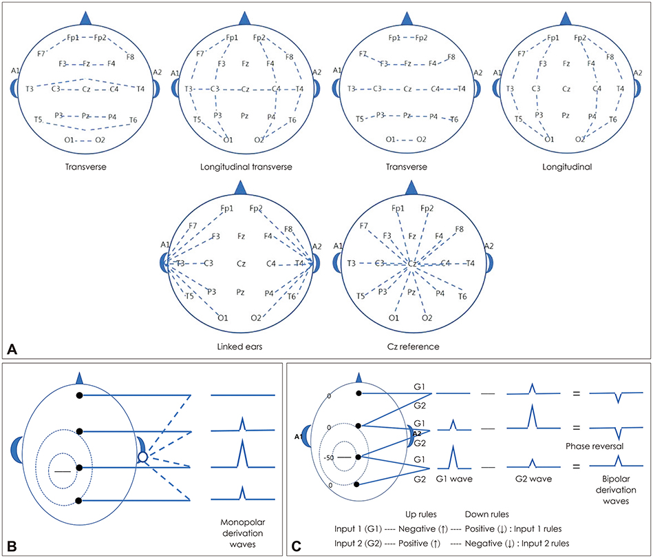
Fig. 1 Montage maps and polarity of EEG. Spectral peaks montage maps. Lines correspond to subtractions used to calculate spectral peaks (A). Monopolar derivation of EEG (B). Bipolar derivation of EEG and phase reversal (C). G1 : Input terminal 1, G2 : Input terminal 2, EEG : Electoencephalography.
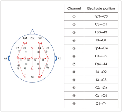
Fig. 2 International 10–20 system and electrode placement for neonatal recording (modified). The International 10–20 system and electrode placement modified for neonates. Some laboratories use an alternative location for the position of the frontal polar electrodes. The ‘Fp3’ is located halfway between the locations of FP1 and F3. The ‘Fp4’ is halfway between the positions of FP2 and F4. Note also that not all laboratories utilize the Pz electrode.34344)

Fig. 3 Modified combinatorial nomenclature for the 10–10 system. Adapted from Acharya et al. J Clin Neurophysiol 2016;33:308–311.47)

Fig. 4 International 10–20 electoencephalography system.7)
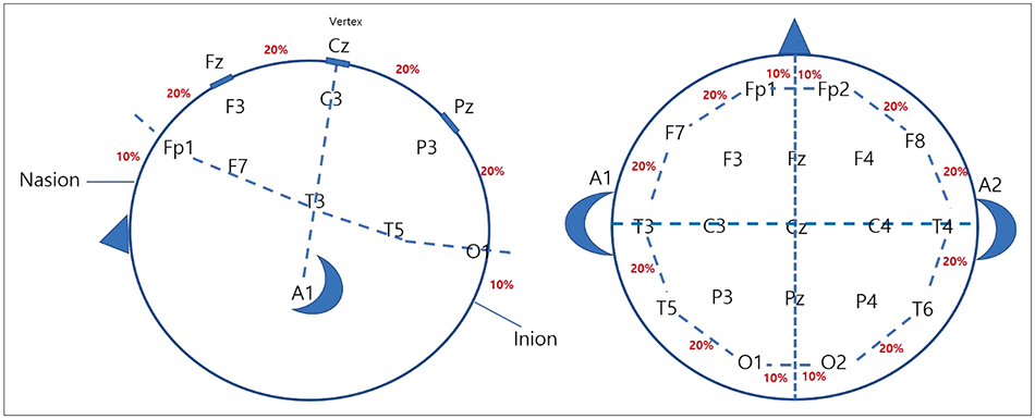
Fig. 5 Electrode placements by international 10–20 electoencephalography system.
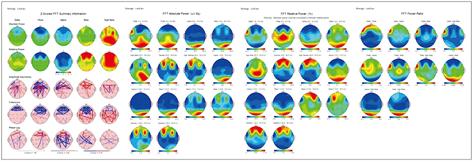
Fig. 6 An example of analysis result of quantitative electroencephalography.

Fig. 7 Examples of normal and abnormal EEG. EEG : Electoencephalography.
Reference
-
1. Shomer DL, Lopes da Silva FH. Niedermyer's electroencephalography: basic principles, clinical applications, and related fields. 7th ed. New York: Oxford University Press;2018.2. Schaul N. The fundamental neural mechanisms of electroencephalography. Electroencephalogr Clin Neurophysiol. 1998; 106:101–107.
Article3. Niedermyer E, Lopes da silva FH. Electroencephalography: basic principles, clinical applications, and related fields. 5th ed. Philadelphia: London: Lippincott Williams & Wilkins;2005.4. William OT. Handbook of EEG interpretation. 2nd ed. New York: New York;2014.5. Chernecky CC, Berger BJ. Laboratory tests and diagnostic procedures. 6th ed. St. Louis: Elsevier;2013.6. Scott NK. Infection prevention: 2013 review and update for neurodiagnostic technologists. Neurodiagn J. 2013; 53:271–288.7. Jasper HH. The 10–20 electrode system of the international federation. Electroencephalogr Clin Neurophysiol. 1958; 10:371–375.8. Sinha SR, Sullivan L, Sabau D, San-Juan D, Dombrowski KE, Halford JJ, et al. American clinical neurophysiology society guideline 1: minimum technical requirements for performing clinical electroencephalography. J Clin Neurophysiol. 2016; 33:303–307.
Article9. Anonymous. Recommendations for the practice of clinical neurophysiology: guidelines of the international federation of clinical neurophysiology. Electroencephalogr Clin Neurophysiol Suppl. 1999; 52:1–304.10. O'Leary JL, Goldring S. Science and epilepsy-Neuroscience gains in epilepsy research. New York: Raven Press;1976.11. Caton R. The electric currents of the brain. Br Med J. 1875; 2:278.
Article12. Coenen A, Fine E, Zayachkivska O. Adolf Beck: a forgotten pioneer in electroencephalography. J Hist Neurosci. 2014; 23:276–286.
Article13. Pravdich-Neminsky W. Ein versuch der registrierung der elektrischen gehirnerscheinungen. Zentralbl Physiol. 1912; 27:951–960.14. Cybulski N, Jeleńska-Macieszyna S. Action currents of the cerebral cortex. Bull Acad Sci Cracov. 1914; 776–781.15. Grzybowski A, Pietrzak K. Napoleon Cybulski (1854–1919). J Neurol. 2013; 260:2942–2943.
Article16. Haas LF. Hans Berger (1873–1941), Richard Caton (1842–1926), and electroencephalography. J Neurol Neurosurg Psychiatry. 2003; 74:9.
Article17. Adrian ED, Matthews BH. The Berger rhythm: potential changes from the occipital lobes in man. Brain. 1934; 57:355–385.
Article18. Grass AM. The electroencephalographic heritage until 1960. Am J EEG Technol. 1984; 24:133–173.
Article19. Berger H. Über das elektrenkephalogramm des menschen. Eur Arch Psychiatry Clin Neurosci. 1929; 87:527–570.
Article20. Britton JW, Frey LC, Hopp JL, Korb P, Koubeissi MZ, Lievens WE, et al. Electroencephalography (EEG): an introductory text and atlas of normal and abnormal findings in adults, children, and infants. Chicago: American Epilepsy Society;2016. p. 1–95.21. Acns.org [homepage on the Internet]. Milwaukee: American Clinical Neurophysiology Society;updated 2017 Jun 14. cited 2019 Mar 8. Available from: https://www.acns.org/about-acns/history.22. National Academy of Engineering. Franklin FO. Biography & autobiography memorial tributes Franklin F. Offner Washington, DC: The National Academies Press;2002. 10:p. 188–192.23. Klem GH, Lüders HO, Jasper HH, Elger C. The ten-twenty electrode system of the international federation. The international federation of clinical neurophysiology. Electroencephalogr Clin Neurophysiol Suppl. 1999; 52:3–6.24. Bozinovski Bozinovski, Bozinovska L, Setakov M. Mobile robot control using alpha wave from the human brain. Zagreb: Proc. Symp. JUREMA;1988. p. 247–249.25. Sutter R, Stevens RD, Kaplan PW. Continuous electroencephalographic monitoring in critically ill patients: indications, limitations, and strategies. Crit Care Med. 2013; 41:1124–1132.
Article26. Nield D. Scientists have connected the brains of 3 people, enabling them to share thoughts. ScienceAlert. 2018; 10. 05.27. Kondraske GV. Neurophysiological measurements. Biomedical Engineering and Instrumentation. 1986; 138–179.28. Fisch BJ, Spehlmann R. EG primer: basic principles of digital and analog EEG. 3rd ed. Amsterdam: Elsevier Science;1999.29. Joseph DB. Medical devices and system: biomedical engineering handbook. 3rd ed. Boca Raton: Informa;2006.30. Teplan M. Fundamentals of EEG measurement. Measurement Science Review. 2002; 2:1–11.31. Fonseca C, Silva Cunha JP, Martins RE, Ferreira VM, Marques de Sá JP, Barbosa MA, et al. A novel dry active electrode for EEG recording. IEEE Trans Biomed Eng. 2007; 54:162–165.
Article32. Egi.com [homepage on the Internet]. Eugene: EGI, A Philips Company;updated 2007 Jan 31. cited 2019 Mar 8. Available from: https://www.egi.com/knowledge-center.33. Speckmann E, Elger CE. Introduction to the neurophysiological basis of the EEG and DC potentials. In : Niedermeyer E, editor. Electroencephalography. Basic principles, clinical applications and related fields. Baltimore: Williams & Wilkins;1999. p. 15–27.34. Ebersole JS. Noninvasive localization of epileptogenic foci by EEG source modeling. Epilepsia. 2000; 41:Suppl 3. S24–S33.
Article35. Buzsaki G, Traub RD, Pedley TA. The cellular basis of EEG activity. In : Ebersole JS, Pedley TA, editors. Current practice of clinical electroencephalography. 3rd ed. Philadelphia: Lippincott Williams & Wilkins;2003. p. 1–11.36. Ebersole JS. Defining epileptogenic foci: past, present, future. J Clin Neurophysiol. 1997; 14:470–483.
Article37. Hong SB, Jung KY. Basic electrophysiology of the electroencephalography. J Korean Neurol Assoc. 2003; 21:225–238.38. Wong PK. Source modelling of the rolandic focus. Brain Topogr. 1991; 4:105–112.
Article39. Bear MF, Connors BW, Paradiso MA. Neuroscience exploring the brain. Baltimore: Lipponcott Williams & Wilkins;2001. p. 606–636.40. Acharya JN, Hani AJ, Thirumala PD, Tsuchida TN. American clinical neurophysiology society guideline 3: a proposal for standard montages to be used in clinical EEG. J Clin Neurophysiol. 2016; 33:312–316.
Article41. Nunez PL, Pilgreen KL. The spline-Laplacian in clinical neurophysiology: a method to improve EEG spatial resolution. J Clin Neurophysiol. 1991; 8:397–413.42. Hrachovy RA, Mizrahi EM. Atlas of neonatal electroencephalography. New York: Demos Medical Publishing;2016. p. 7–13.43. Clancy RR, Bergqvist AGC, Dlugos DJ. Neonatal electroencephalography. In : Ebersole JS, Pedley TA, editors. Current practice of clinical electroencephalography. Philadelphia: Lippincott Williams & Wilkins;2003. p. 160–234.44. Shellhaas RA, Chang T, Tsuchida T, Scher MS, Riviello JJ, Abend NS, et al. The American Clinical Neurophysiology Society's guideline on continuous electroencephalography monitoring in neonates. J Clin Neurophysiol. 2011; 28:611–617.
Article45. Kuratani J, Pearl PL, Sullivan L, Riel-Romero RM, Cheek J, Stecker M, et al. American Clinical Neurophysiology Society guideline 5: minimum technical standards for pediatric electroencephalography. J Clin Neurophysiol. 2016; 33:320–323.
Article46. Tsuchida TN, Acharya JN, Halford JJ, Kuratani JD, Sinha SR, Stecker MM, et al. American Clinical Neurophysiology Society: EEG guidelines introduction. J Clin Neurophysiol. 2016; 33:301–302.
Article47. Acharya JN, Hani A, Cheek J, Thirumala P, Tsuchida TN. American Clinical Neurophysiology Society guideline 2: guidelines for standard electrode position nomenclature. J Clin Neurophysiol. 2016; 33:308–311.
Article48. Koo DL, Kim WJ, Lee SA, Kim JM, Kim J, Park S. Fundamental requirements for performing electroencephalography. Ann Clin Neurophysiol. 2017; 19:113–117.
Article49. Barry W, Jones GM. Influence of eye lid movement upon electro-oculographic recording of vertical eye movements. Aerosp Med. 1965; 36:855–858.50. Iwasaki M, Kellinghaus C, Alexopoulos AV, Burgess RC, Kumar AN, Han YH, et al. Effects of eyelid closure, blinks, and eye movements on the electroencephalogram. Clin Neurophysiol. 2005; 116:878–885.
Article51. Keren AS, Yuval-Greenberg S, Deouell LY. Saccadic spike potentials in gamma-band EEG: characterization, detection and suppression. Neuroimage. 2010; 49:2248–2263.
Article52. Epstein CM, Andriola MR. Introduction to EEG and evoked potentials. New York: Lippincott;1983.53. Symeonidou ER, Nordin AD, Hairston WD, Ferris DP. Effects of cable sway, electrode surface area, and electrode mass on electroencephalography signal quality during motion. Sensors (Basel). 2018; 18:E1073.
Article54. Oliveira AS, Schlink BR, Hairston WD, König P, Ferris DP. Induction and separation of motion artifacts in EEG data using a mobile phantom head device. J Neural Eng. 2016; 13:036014.
Article55. Jung TP, Makeig S, Humphries C, Lee TW, McKeown MJ, Iragui V, et al. Removing electroencephalographic artifacts by blind source separation. Psychophysiology. 2000; 37:163–178.
Article56. Joyce CA, Gorodnitsky IF, Kutas M. Automatic removal of eye movement and blink artifacts from EEG data using blind component separation. Psychophysiology. 2004; 41:313–325.
Article57. Shackman AJ, McMenamin BW, Maxwell JS, Greischar LL, Davidson RJ. Identifying robust and sensitive frequency bands for interrogating neural oscillations. Neuroimage. 2010; 51:1319–1333.
Article58. Nolan H, Whelan R, Reilly RB. FASTER: fully automated statistical thresholding for EEG artifact rejection. J Neurosci Methods. 2010; 192:152–162.
Article59. Fitzgibbon SP, Lewis TW, Powers DM, Whitham EW, Willoughby JO, Pope KJ. Surface laplacian of central scalp electrical signals is insensitive to muscle contamination. IEEE Trans Biomed Eng. 2013; 60:4–9.
Article60. Kondylis ED, Wozny TA, Lipski WJ, Popescu A, DeStefino VJ, Esmaeili B, et al. Detection of high-frequency oscillations by hybrid depth electrodes in standard clinical intracranial EEG recordings. Front Neurol. 2014; 5:149.
Article61. Hämäläinen M, Hari R, Ilmoniemi RJ, Knuutila J, Lounasmaa OV. Magnetoencephalography—theory, instrumentation, and applications to noninvasive studies of the working human brain. Rev Mod Phys. 1993; 65:413–497.
Article62. Murakami S, Okada Y. Contributions of principal neocortical neurons to magnetoencephalography and electroencephalography signals. J Physiol. 2006; 575:925–936.
Article63. Nuwer M. Assessment of digital EEG, quantitative EEG, and EEG brain mapping: report of the American Academy of neurology and the American Clinical Neurophysiology Society. Neurology. 1997; 49:277–292.
Article64. Duffy FH, Hughes JR, Miranda F, Bernad P, Cook P. Status of quantitative EEG (QEEG) in clinical practice, 1994. Clin Electroencephalogr. 1994; 25:VI–XXII.
Article65. Chung HJ, Hur YJ. Diagnosis of neonatal seizures. Korean J Pediatr. 2009; 52:964–970.
Article66. Karl FS. The aEEG booklet a quick overview for the practical routine. 1st ed. Tokyo: Nihon Kohden Corporation;2012.67. Maynard D, Prior PF, Scott DF. Device for continuous monitoring of cerebral activity in resuscitated patients. Br Med J. 1969; 4:545–546.
Article68. Azzopardi DT. Cerebral function monitoring: addition to CFM handbook for users of the olympic CFM 6000. London: Imperial College London;2004.69. Anderson J. Cognitive psychology and its implications. 6th ed. New York: Worth;2004. p. 17.70. Nunez PL, Srinivasan R. Electric fields of the brain: the neurophysics of EEG. Oxford: Oxford;1981.71. Creutzfeldt OD, Watanabe S, Lux HD. Relations between EEG phenomena and potentials of single cortical cells. I. Evoked responses after thalamic and erpicortical stimulation. Electroencephalogr Clin Neurophysiol. 1966; 20:1–18.
Article72. Tanzer OI. Numerical modeling in electro- and magnetoencephalography [dissertation]. Espoo: Helsinki University of Technology;2006.73. Tatum WO, Ellen R. Grass lecture: extraordinary EEG. Neurodiagn J. 2014; 54:3–21.74. Cahn BR, Polich J. Meditation states and traits: EEG, ERP, and neuroimaging studies. Psychol Bull. 2006; 132:180–211.
Article75. Gerrard P, Malcolm R. Mechanisms of modafinil: a review of current research. Neuropsychiatr Dis Treat. 2007; 3:349–364.76. Niedermeyer E. Alpha rhythms as physiological and abnormal phenomena. Int J Psychophysiol. 1997; 26:31–49.
Article77. Feshchenko VA, Reinsel RA, Veselis RA. Multiplicity of the alpha rhythm in normal humans. J Clin Neurophysiol. 2001; 18:331–344.78. Pfurtscheller G, Lopes da Silva FH. Event-related EEG/MEG synchronization and desynchronization: basic principles. Clin Neurophysiol. 1999; 110:1842–1857.
Article79. Oberman LM, Hubbard EM, McCleery JP, Altschuler EL, Ramachandran VS, Pineda JA. EEG evidence for mirror neuron dysfunction in autism spectrum disorders. Brain Res Cogn Brain Res. 2005; 24:190–198.
Article80. Anderson AL, Thomason ME. Functional plasticity before the cradle: a review of neural functional imaging in the human fetus. Neurosci Biobehav Rev. 2013; 37:2220–2232.
Article81. Santamaria J, Chiappa KH. The EEG of drowsiness in normal adults. J Clin Neurophysiol. 1987; 4:327–382.
Article82. Frohlich J, Senturk D, Saravanapandian V, Golshani P, Reiter LT, Sankar R, et al. A quantitative electrophysiological biomarker of duplication 15q11.2-q13.1 syndrome. PLoS One. 2016; 11:e0167179.
Article83. Kisley MA, Cornwell ZM. Gamma and beta neural activity evoked during a sensory gating paradigm: effects of auditory, somatosensory and cross-modal stimulation. Clin Neurophysiol. 2006; 117:2549–2563.
Article84. Kanayama N, Sato A, Ohira H. Crossmodal effect with rubber hand illusion and gamma-band activity. Psychophysiology. 2007; 44:392–402.
Article85. Gastaut H. Electrocorticographic study of the reactivity of rolandic rhythm. Rev Neurol (Paris). 1952; 87:176–182.86. Krishnan V, Chang BS, Schomer DL. Normal EEG in wakefulness and sleep: adult and elderly. In : Schomer DL, Lopes da Silva FH, editors. Niedermyer's electroencephalography;basic principles, clinical applications, and related fields. New York: Oxford;2018. p. 202–228.87. Montez T, Poil SS, Jones BF, Manshanden I, Verbunt JP, Van Dijk BW, et al. Altered temporal correlations in parietal alpha and prefrontal theta oscillations in early-stage Alzheimer disease. Proc Natl Acad Sci U S A. 2009; 106:1614–1619.
Article88. Scher MS. Electroencephalography of the newborn. In : Niedermeyer E, Da Silva FH, editors. Electroencephalography: basic principles, clinical applications, and related fields. Philadelphia: Lippincott Williams & Wilkins;2005. p. 937–990.89. Kellaway P. Ordely approach to visual analysis: elements of the normal EEG and their characteristics in children and adults. In : Ebersole JS, Pedley TA, editors. Current practice of clinical electroencephalography. Philadelphia: Lippincott Williams & Wilkins;2003. p. 100–159.90. Clancy RR, Bergqvist AGC, Dlugos DJ, Nordli D. Normal Pediatric EEG: Neonates and Children. In : Ebersole JS, Husain AM, Nordli AR, editors. Current practice of clinical electroencephalography. Philadelphia: Wolters Kluwer;2014. p. 125–212.91. Obeid R, Tsuchida TN. Treatment effects on neonatal EEG. J Clin Neurophysiol. 2016; 33:376–381.
Article92. Francis IC, Loughhead JA. Bell's phenomenon: a study of 508 patients. Aust J Ophthalmol. 1984; 12:15–21.
Article93. Markand ON. Alpha rhythms. J Clin Neurophysiol. 1990; 7:163–189.
Article94. Clarke AR, Barry RJ, Dupuy FE, McCarthy R, Selikowitz M, Johnstone SJ. Excess beta activity in the EEG of children with attention-deficit/hyperactivity disorder: a disorder of arousal? Int J Psychophysiol. 2013; 89:314–319.
Article95. Brigo F. Lambda waves. Am J Electroneurodiagnostic Technol. 2011; 51:105–113.
Article96. Daly DD, Pedley TA. Current practice of clinical electroencephalography. 2nd ed. Philadelphia: Lippincott Williams & Wilkins;1997.97. Shanbao T, Nitish V. Thankor. Quantitative EEG analysis methods and applications. Norwood: Artech House;2009.98. Tatum WO, Olga S, Ochoa JG, Munger Clary H, Cheek J, Drislane F, et al. American clinical neurophysiology society guideline 7: guidelines for EEG reporting. J Clin Neurophysiol. 2016; 33:328–332.
Article99. Kaplan PW, Benbadis SR. How to write an EEG report: dos and don'ts. Neurology. 2013; 80:S43–S46.
Article100. Halford JJ, Sabau D, Drislane FW, Tsuchida TN, Sinha SR. American clinical neurophysiology society guideline 4: recording clinical EEG on digital media. J Clin Neurophysiol. 2016; 33:317–319.
Article
- Full Text Links
-
- Actions
-
Cited
- CITED
-
- Close
- Share
-
- Similar articles
-
- Introduction to Starting Upper Gastrointestinal Endoscopy: Proper Insertion, Complete Observation, and Appropriate Photographing
- The Clinical Use of Electroencephalography in Psychiatry
- The Significance of Electroencephalography in Neonate and Prematurity
- Addiction and Quantitative Electroencephalography
- Back to the Basics!
