Obstet Gynecol Sci.
2017 Jul;60(4):329-335. 10.5468/ogs.2017.60.4.329.
Second trimester cervical length measurement for prediction spontaneous preterm birth in an unselected risk population
- Affiliations
-
- 1Mario Palmério University Hospital, University of Uberaba (UNIUBE), Uberaba-MG, Brazil.
- 2Department of Obstetrics and Gynecology, Ribeirão Preto Medical School, University of São Paulo (FMRP-USP), Ribeirão Preto-SP, Brazil.
- 3Department of Obstetrics and Gynaecology, Monash University Faculty of Medicine Nursing and Health Sciences, Clayton, Victoria, Australia.
- 4Department of Obstetrics, Paulista School of Medicine, Federal University of São Paulo (EPM-UNIFESP), São Paulo-SP, Brazil. araujojred@terra.com.br
- KMID: 2386273
- DOI: http://doi.org/10.5468/ogs.2017.60.4.329
Abstract
OBJECTIVE
To assess the predictive capacity of cervical length (CL) measurement underwent during the second trimester ultrasound for prediction preterm birth <32, 34, and 37 weeks of gestation in an unselected risk population.
METHODS
A retrospective cohort study was performed with 751 singleton pregnancies between 20 and 24+6 weeks of gestation. The CL measurement (mm) using the transvaginal route was obtained in a sagittal view and the calipers positioned to measure the linear distance between the triangular area of echodensity at the external os and the internal os. To compare the preterm (<37 weeks) and term births (≥37 weeks), we used unpaired t test. We assessed whether the CL measurement was dependent of gestational age by performing a linear regression and assessing the coefficient of determination (R²). We additionally assessed the accuracy of CL measurement to predict preterm birth by assessing the area under receiver operating characteristics curves with its respective confidence intervals (CIs) 95%.
RESULTS
Preterm birth <37 weeks was found in 13.6% (102/751) of pregnant women. Short cervix (≤25 mm) was found in 2.7% (20/751) of pregnancies. Only 30% (6/20) of pregnant women with short cervix have used progesterone to prevent preterm birth. There was a weak correlation between CL measurement and gestational age at delivery (R²=0.01, P=0.002). Receiver operating characteristics curve analysis of the ability of CL measurement to predict preterm birth <32, 34, and 37 weeks, showed an area under the curve of 0.693 (95% CI, 0.512 to 0.874), 0.472 (95% CI, 0.353 to 0.591), 0.490 (95% CI, 0.426 to 0.555), respectively.
CONCLUSION
There was a weak correlation between CL measurement and gestational age at delivery. In an unselected population, CL measurement screening at 20 to 24+6 weeks of gestation does not seem to be a good predictor of preterm birth.
Keyword
MeSH Terms
Figure
-
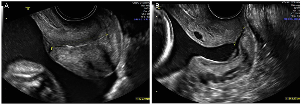
Fig. 1 Transvaginal ultrasound examination showing the cervical length measurement at 22 weeks of gestation. (A) Normal cervical length measurement and (B) short cervix with funneling.
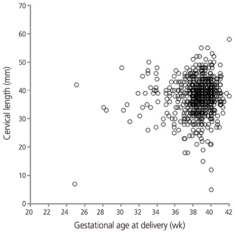
Fig. 2 Scatter plot of the correlation between cervical length measurement (mm) and gestational age (weeks) at delivery.

Fig. 3 Receiver operating characteristics curves to display the cervical length measurement ability to predict preterm birth (A) <32, (B) 34, and (C) 37 weeks of gestation. Diagonal segments are produced by ties.
Reference
-
1. Iams JD, Romero R, Culhane JF, Goldenberg RL. Primary, secondary, and tertiary interventions to reduce the morbidity and mortality of preterm birth. Lancet. 2008; 371:164–175.2. Manuck TA, Rice MM, Bailit JL, Grobman WA, Reddy UM, Wapner RJ, et al. Preterm neonatal morbidity and mortality by gestational age: a contemporary cohort. Am J Obstet Gynecol. 2016; 215:103.3. Heath VC, Southall TR, Souka AP, Elisseou A, Nicolaides KH. Cervical length at 23 weeks of gestation: prediction of spontaneous preterm delivery. Ultrasound Obstet Gynecol. 1998; 12:312–317.4. Miller ES, Tita AT, Grobman WA. Second-trimester cervical length screening among asymptomatic women: an evaluation of risk-based strategies. Obstet Gynecol. 2015; 126:61–66.5. Fonseca EB, Celik E, Parra M, Singh M, Nicolaides KH. Fetal Medicine Foundation Second Trimester Screening Group. Progesterone and the risk of preterm birth among women with a short cervix. N Engl J Med. 2007; 357:462–469.6. Cahill AG, Odibo AO, Caughey AB, Stamilio DM, Hassan SS, Macones GA, et al. Universal cervical length screening and treatment with vaginal progesterone to prevent preterm birth: a decision and economic analysis. Am J Obstet Gynecol. 2010; 202:548.7. Sonek J, Shellhaas C. Cervical sonography: a review. Ultrasound Obstet Gynecol. 1998; 11:71–78.8. Sotiriadis A, Papatheodorou SI, Martins WP. Synthesizing Evidence from Diagnostic Accuracy TEsts: the SEDATE guideline. Ultrasound Obstet Gynecol. 2016; 47:386–395.9. Hamilton BE, Martin JA, Ventura SJ. Births: preliminary data for 2006. Natl Vital Stat Rep. 2007; 56:1–18.10. Leal MD, Esteves-Pereira AP, Nakamura-Pereira M, Torres JA, Theme-Filha M, Domingues RM, et al. Prevalence and risk factors related to preterm birth in Brazil. Reprod Health. 2016; 13:Suppl 3. 127.11. Romero R, Nicolaides KH, Conde-Agudelo A, O'Brien JM, Cetingoz E, Da Fonseca E, et al. Vaginal progesterone decreases preterm birth ≤ 34 weeks of gestation in women with a singleton pregnancy and a short cervix: an updated meta-analysis including data from the OPPTIMUM study. Ultrasound Obstet Gynecol. 2016; 48:308–317.12. Society for Maternal-Fetal Medicine (SMFM). McIntosh J, Feltovich H, Berghella V, Manuck T. The role of routine cervical length screening in selected high- and low-risk women for preterm birth prevention. Am J Obstet Gynecol. 2016; 215:B2–B7.13. Temming LA, Durst JK, Tuuli MG, Stout MJ, Dicke JM, Macones GA, et al. Universal cervical length screening: implementation and outcomes. Am J Obstet Gynecol. 2016; 214:523.14. Son M, Grobman WA, Ayala NK, Miller ES. A universal mid-trimester transvaginal cervical length screening program and its associated reduced preterm birth rate. Am J Obstet Gynecol. 2016; 214:365.15. Hassan SS, Romero R, Vidyadhari D, Fusey S, Baxter JK, Khandelwal M, et al. Vaginal progesterone reduces the rate of preterm birth in women with a sonographic short cervix: a multicenter, randomized, double-blind, placebo-controlled trial. Ultrasound Obstet Gynecol. 2011; 38:18–31.16. Peixoto AB, da Cunha Caldas TM, Alamy AH, Martins WP, Bruns RF, Araujo Junior E. Reference values for the cervical length measurement in the second trimester of pregnancy using the transvaginal ultrasound in a large Brazilian population. Obstet Gynecol Sci. 2016; 59:303–306.17. Hughes K, Kane SC, Araujo Junior E, Da Silva Costa F, Sheehan PM. Cervical length as a predictor for spontaneous preterm birth in high-risk singleton pregnancy: current knowledge. Ultrasound Obstet Gynecol. 2016; 48:7–15.18. Celik E, To M, Gajewska K, Smith GC, Nicolaides KH. Fetal Medicine Foundation Second Trimester Screening Group. Cervical length and obstetric history predict spontaneous preterm birth: development and validation of a model to provide individualized risk assessment. Ultrasound Obstet Gynecol. 2008; 31:549–554.
- Full Text Links
-
- Actions
-
Cited
- CITED
-
- Close
- Share
-
- Similar articles
-
- Cervical Length Measurement to Predict Preterm Birth: Clinical Application and Treatment Modalities
- How to screen the cervix and reduce the risk of spontaneous preterm birth in asymptomatic women without a prior preterm birth
- Maternal Characteristics, Short Mid-Trimester Cervical Length, and Preterm Delivery
- Pregnancy outcome after cervical conization: risk factors for preterm delivery and the efficacy of prophylactic cerclage
- Uterine Cervical Length Measurement by Transvaginal Ultrasonography for Prediction of Preterm Delivery
