Ann Dermatol.
2011 Nov;23(4):508-511.
Benign Cephalic Histiocytosis: A Case Report
- Affiliations
-
- 1Department of Dermatology, Faculty of Medicine, Zonguldak Karaelmas University, Zonguldak, Turkey. rafkoca@yahoo.com
- 2Department of Pathology, Faculty of Medicine, Zonguldak Karaelmas University, Zonguldak, Turkey.
- 3Department of Dermatology, Selcuklu Medical Faculty, Selcuk University, Konya, Turkey.
Abstract
- Histiocytic skin disorders are usually classified as either Langerhans' cell histiocytosis (LCH) or non LCH, based on the pathology. Benign cephalic histiocytosis (BCH) is a rare type of non-Langerhans histiocytitic disorder and is characterized by self-healing multiple small eruptions of yellow to red-brown papules on the face and upper trunk. Histologic features of this disorder show dermal proliferation of histiocytes that have intracytoplasmic comma-shaped bodies, coated vesicles and desmosome-like structures. In this study, we report on a 7-month-old boy who contained small yellow-red papules on his face that spread to his upper trunk. The clinical and histologic features in this patient were consistent with BCH.
Keyword
Figure
-
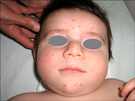
Fig. 1 Small yellow-red papules scattered on the face, forehead and chin.
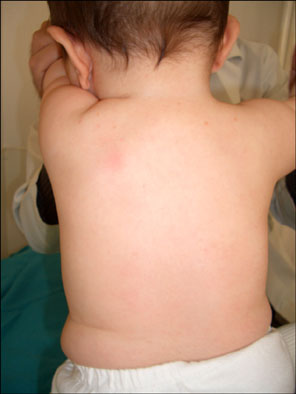
Fig. 2 Discrete small yellow-red papules seen on the upper back.
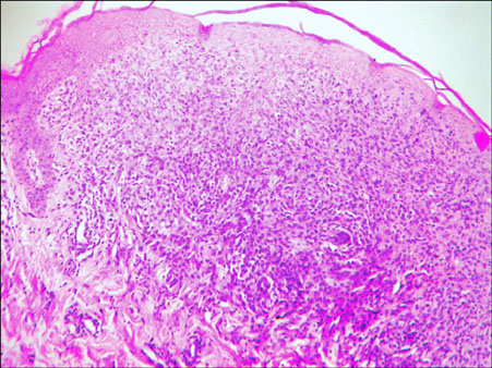
Fig. 3 Proliferations of pleomorphic epitheliod histiocytic cells within the upper- and mid-dermis (H&E, original magnification ×100).
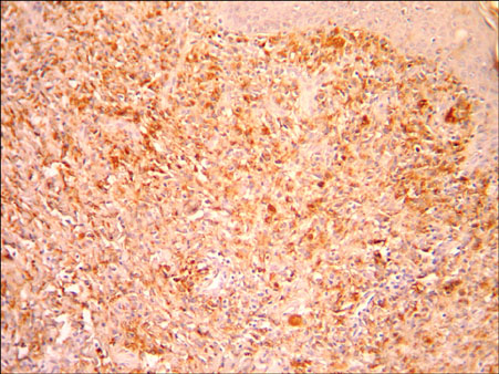
Fig. 4 Immunohistochemistry was positive stained for CD68 (biotin-streptavidin peroxidase system, DAB, original magnification ×200).
Reference
-
1. Gianotti F, Caputo R, Ermacora E. Singular "infantile histiocytosis with cells with intracytoplasmic vermiform particles". Bull Soc Fr Dermatol Syphiligr. 1971. 78:232–233.2. Jih DM, Salcedo SL, Jaworsky C. Benign cephalic histiocytosis: a case report and review. J Am Acad Dermatol. 2002. 47:908–913.
Article3. Zelger BW, Sidoroff A, Orchard G, Cerio R. Non-Langerhans cell histiocytoses. A new unifying concept. Am J Dermatopathol. 1996. 18:490–504.4. Dadzie O, Hopster D, Cerio R, Wakeel R. Benign cephalic histiocytosis in a British-African child. Pediatr Dermatol. 2005. 22:444–446.
Article5. Hasegawa S, Deguchi M, Chiba-Okada S, Aiba S. Japanese case of benign cephalic histiocytosis. J Dermatol. 2009. 36:69–71.
Article6. Baler JS, DiGregorio FM, Hashimoto K. Facial papules in a child. Benign cephalic histiocytosis. Arch Dermatol. 1995. 131:610–611.
Article7. Gianotti F, Caputo R, Ermacora E, Gianni E. Benign cephalic histiocytosis. Arch Dermatol. 1986. 122:1038–1043.
Article8. Sidwell RU, Francis N, Slater DN, Mayou SC. Is disseminated juvenile xanthogranulomatosis benign cephalic histiocytosis. Pediatr Dermatol. 2005. 22:40–43.
Article9. Watabe H, Soma Y, Matsutani Y, Baba T, Mizoguchi M. Case 2: benign cephalic histiocytosis. Clin Exp Dermatol. 2002. 27:341–342.10. Saez-De-Ocariz M, Lopez-Corella E, Duran-McKinster C, Orozco-Covarrubias L, Ruiz-Maldonado R. Benign cephalic histiocytosis preceding the development of insulindependent diabetes mellitus. Pediatr Dermatol. 2006. 23:101–102.
Article11. Weston WL, Travers SH, Mierau GW, Heasley D, Fitzpatrick J. Benign cephalic histiocytosis with diabetes insipidus. Pediatr Dermatol. 2000. 17:296–298.12. Gianotti R, Alessi E, Caputo R. Benign cephalic histiocytosis: a distinct entity or a part of a wide spectrum of histiocytic proliferative disorders of children? A histopathological study. Am J Dermatopathol. 1993. 15:315–319.13. Caputo R, Ermacora E, Gelmetti C, Berti E, Gianni E, Nigro A. Generalized eruptive histiocytoma in children. J Am Acad Dermatol. 1987. 17:449–454.
Article14. Winkelmann RK, Kossard S, Fraga S. Eruptive histiocytoma of childhood. Arch Dermatol. 1980. 116:565–570.
Article15. Rodriguez-Jurado R, Duran-McKinster C, Ruiz-Maldonado R. Benign cephalic histiocytosis progressing into juvenile xanthogranuloma: a non-Langerhans cell histiocytosis transforming under the influence of a virus? Am J Dermatopathol. 2000. 22:70–74.
Article16. Zelger BG, Zelger B, Steiner H, Mikuz G. Solitary giant xanthogranuloma and benign cephalic histiocytosis--variants of juvenile xanthogranuloma. Br J Dermatol. 1995. 133:598–604.
Article17. Aliaga A, Alegre VA, Hernández M, Febrer MI, Martinez A, Portea JM. Benign cephalic histiocytosis vs generalized eruptive histiocytoma. J Cutan Pathol. 1989. 16:293–296.18. Goday JJ, Raton JA, Landa N, Eizaguirre X, Diaz-Pérez JL. Benign cephalic histiocytosis--study of a case. Clin Exp Dermatol. 1993. 18:280–282.
Article19. Umbert IJ, Winkelmann RK. Eruptive histiocytoma. J Am Acad Dermatol. 1989. 20:958–964.
Article
