Ann Dermatol.
2011 Sep;23(Suppl 1):S16-S19. 10.5021/ad.2011.23.S1.S16.
A Case of Benign Cephalic Histiocytosis
- Affiliations
-
- 1Department of Dermatology, College of Medicine, Hallym University, Anyang, Korea. dermakkh@yahoo.co.kr
- 2Department of Dermatology, College of Medicine, Hallym University, Chuncheon, Korea.
- KMID: 2156742
- DOI: http://doi.org/10.5021/ad.2011.23.S1.S16
Abstract
- Benign cephalic histiocytosis (BCH) is a rare non-Langerhans cell histiocytosis of unknown etiology. Clinically, lesions are characterized by small, red-to-yellow papules distributed mainly on the head, face, neck, and shoulders of infants and children. Histopathological specimens show massive histiocytic infiltration of the superficial dermis. Immunohistochemically, they are positive for CD68, but negative for CD1a and S-100. Two cases have been reported so far in the relevant work published in Korean literature. Herein, we report on an additional case of BCH.
Keyword
Figure
-
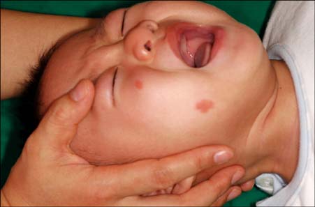
Fig. 1 Erythematous grouped papules on wheal-like plaques on the right cheek.
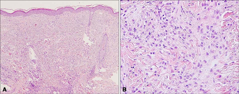
Fig. 2 (A) Diffuse cell infiltrates were observed throughout the dermis (H&E, ×100). (B) High power view demonstrating proliferation of large, epithelioid histiocytic cells with eosinophilic cytoplasm (H&E, ×400).
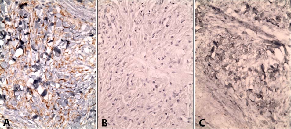
Fig. 3 Immunohistochemical staining shows (A) positive for CD68 (×400), negative for (B) S-100 and (C) CD1a (×400).
Reference
-
1. Gianotti F, Caputo R, Ermacora E. Singular "infantile histiocytosis with cells with intracytoplasmic vermiform particles". Bull Soc Fr Dermatol Syphiligr. 1971. 78:232–233.2. Kim DH, Bang DS, Han SW, Whang KC, Lee HE. A case of benign cephalic histiocytosis. Korean J Dermatol. 1986. 24:433–438.3. Jeong JS, Jang EJ, Lee JY, Suh KS, Yoon TY. A case of benign cephalic histiocytosis. Korean J Dermatol. 2008. 46:1292–1295.4. Barsky BL, Lao I, Barsky S, Rhee HL. Benign cephalic histiocytosis. Arch Dermatol. 1984. 120:650–655.
Article5. Jih DM, Salcedo SL, Jaworsky C. Benign cephalic histiocytosis: a case report and review. J Am Acad Dermatol. 2002. 47:908–913.
Article6. Zelger BG, Zelger B, Steiner H, Mikuz G. Solitary giant xanthogranuloma and benign cephalic histiocytosis--variants of juvenile xanthogranuloma. Br J Dermatol. 1995. 133:598–604.
Article7. Rodriguez-Jurado R, Duran-McKinster C, Ruiz-Maldonado R. Benign cephalic histiocytosis progressing into juvenile xanthogranuloma: a non-Langerhans cell histiocytosis transforming under the influence of a virus? Am J Dermatopathol. 2000. 22:70–74.
Article8. Sidwell RU, Francis N, Slater DN, Mayou SC. Is disseminated juvenile xanthogranulomatosis benign cephalic histiocytosis? Pediatr Dermatol. 2005. 22:40–43.
Article9. Weston WL, Travers SH, Mierau GW, Heasley D, Fitzpatrick J. Benign cephalic histiocytosis with diabetes insipidus. Pediatr Dermatol. 2000. 17:296–298.10. Saez-De-Ocariz M, Lopez-Corella E, Duran-McKinster C, Orozco-Covarrubias L, Ruiz-Maldonado R. Benign cephalic histiocytosis preceding the development of insulin-dependent diabetes mellitus. Pediatr Dermatol. 2006. 23:101–102.
Article11. Watabe H, Soma Y, Matsutani Y, Baba T, Mizoguchi M. Case 2: benign cephalic histiocytosis. Clin Exp Dermatol. 2002. 27:341–342.12. Dadzie O, Hopster D, Cerio R, Wakeel R. Benign cephalic histiocytosis in a British-African child. Pediatr Dermatol. 2005. 22:444–446.
Article13. Hasegawa S, Deguchi M, Chiba-Okada S, Aiba S. Japanese case of benign cephalic histiocytosis. J Dermatol. 2009. 36:69–71.
Article
