Ann Dermatol.
2016 Oct;28(5):643-645. 10.5021/ad.2016.28.5.643.
Cutaneous Amyloidoma: A Rare Case Report
- Affiliations
-
- 1Department of Dermatology and Venereology, Erciyes University Faculty of Medicine, Kayseri, Turkey. demetkartal@hotmail.com
- 2Department of Pathology, Erciyes University Faculty of Medicine, Kayseri, Turkey.
- KMID: 2382894
- DOI: http://doi.org/10.5021/ad.2016.28.5.643
Abstract
- No abstract available.
Figure
-
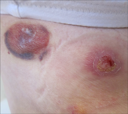
Fig. 1 Two skin-colored firm nodules with a smooth surface, one of them had a hemoragic border and the other was ulcerated.
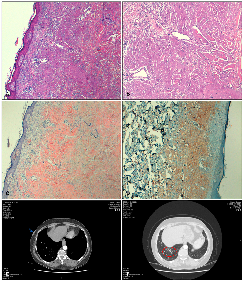
Fig. 2 (A, B) Dermal connective tissue with deposits of amorphous material (H&E). (C) Extensive amyloid deposits in the entire dermis were positive with Congo-red staining. (D) The amyloid type was determined immunohistochemically as AA amyloid. (E) Axial computed tomography (CT) scan at the soft tissue window shows skin lesion (arrow). (F) Axial CT scan at the lung window shows cluster of multiple small nodules in the right lower lobe (circle).
Reference
-
1. Bauer WH, Kuzma JF. Solitary tumors of atypical amyloid (paramyloid). Am J Clin Pathol. 1949; 19:1097–1112.
Article2. Reitboeck JG, Feldmann R, Loader D, Breier F, Steiner A. Primary cutaneous amyloidoma: a case report. Case Rep Dermatol. 2014; 6:264–267.
Article3. Biewend ML, Menke DM, Calamia KT. The spectrum of localized amyloidosis: a case series of 20 patients and review of the literature. Amyloid. 2006; 13:135–142.
Article4. Banno S, Matsumoto Y, Hayami Y, Sugiura Y, Yoshinouchi T, Ueda R. Pulmonary AL amyloidosis in a patient with primary Sjögren syndrome. Mod Rheumatol. 2002; 12:84–88.
Article5. Mlika M, Ayadi-Kaddour A, Marghli A, Ridène I, Maalej S, El Mezni F. A rare pulmonary lesion association. Rev Pneumol Clin. 2012; 68:303–306.
- Full Text Links
-
- Actions
-
Cited
- CITED
-
- Close
- Share
-
- Similar articles
-
- Soft Tissue Amyloidoma of Upper Extremity: A Case Report
- A Case of Bilateral Trigeminal Amyloidoma Diagnosed Through an Endoscopic Transsphenoidal Approach
- Calcific Amyloidoma of Tibialis Anterior Muscle: Case Report
- CT and MRI Findings of Small Bowel Involvement of Amyloidosis Mimicking Small Bowel Polyposis Syndrome: a Case Report
- Spinal Cord Compression by Primary Amyloidoma of the Spine
