Calcific Amyloidoma of Tibialis Anterior Muscle: Case Report
- Affiliations
-
- 1Department of Orthopaedic Surgery, College of Medicine,Chungbuk National University, Cheongju, Korea. ymkim@chungbuk.ac.kr
- 2Department of Pathology, College of Medicine,Chungbuk National University, Cheongju, Korea.
- KMID: 2186455
- DOI: http://doi.org/10.4055/jkoa.2008.43.3.374
Abstract
- Calcific amyloidoma of the soft tissue is quite rare and it is difficult to make a differential diagnosis from other lesions such as osteomyelitis or bone tumor. We encountered a case of a calcified amyloidoma found in the anterior tibial muscle that occurred more than 20 years after a proximal tibial fracture adjacent to the origin of the muscle. The features of the lesion resembled osteomyelitis. Satisfactory result was obtained by a thorough mass excision. We report this case with review of the relevant literature.
Keyword
Figure
-
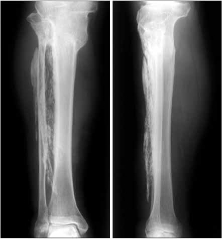
Fig. 1 There were a large amount of high density streaky sclerotic lesions over almost the whole space of the tibialis anterior muscle. An old fracture of the proximal tibia was observed with complete union.

Fig. 2 The sclerotic mass on the plain X-ray was observed as low signal intensity (white arrow) on the T2-weighted axial (A) and sagittal (B) MR images. Overlying subcutaneous tissue showed high signal changes (dark arrow head) suggesting fluid collection.
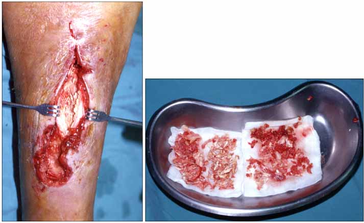
Fig. 3 There were a large amount of sawdust like osseous components around the interosseous membrane, which were scattered in the anterior side of the right tibia.
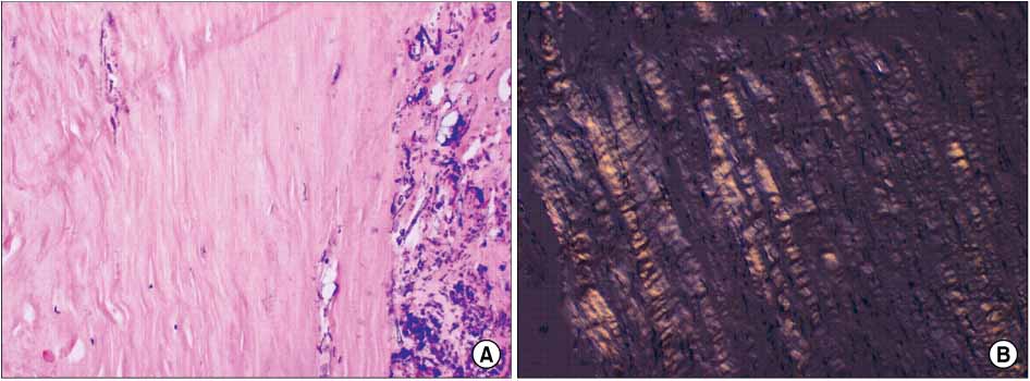
Fig. 4 The lesion appeared as an amorphous, eosinophilic, hyaline, extracellular substance with calcification (A: HE, ×100). Under polarized light, the Congo red-stained amyloid in the tissue showed green birefringence (B, ×100).
Reference
-
1. Bardin RL, Barnes CE, Stanton CA, Geisinger KR. Soft tissue amyloidoma of the extremities: a case report and review of the literature. Arch Pathol Lab Med. 2004. 128:1270–1273.
Article2. Cobby MJ, Adler RS, Swartz R, Martel W. Dialysis-related amyloid arthropathy: MR findings in four patients. AJR Am J Roentgenol. 1991. 157:1023–1027.
Article3. Gean-Marton AD, Kirsch CF, Vezina LG, Weber AL. Focal amyloidosis of the head and neck: evaluation with CT and MR imaging. Radiology. 1991. 181:521–525.
Article4. Glenner GG. Amyloid deposits and amyloidosis. N Engl J Med. 1980. 302:1283–1292.
Article5. Kato H, Toei H, Furuse M, Suzuki K, Hironaka M, Saito K. Primary localized amyloidosis of the urinary bladder. Eur Radiol. 2003. 13:Suppl 6. 109–112.
Article6. Krishnan J, Chu WS, Elrod JP, Frizzera G. Tumoral presentation of amyloidosis (amyloidomas) in soft tissues. A report of 14 cases. Am J Clin Pathl. 1993. 100:135–144.7. Kyle RA, Greipp PR. Amyloidomsis (AL). Clinical and laboratory features in 229 cases. Mayo Clin Proc. 1983. 58:665–683.8. Möllers MJ, van Schaik JP, van der Putte SC. Pulmonary amyloidoma. Histologic proof yielded by transthoracic coaxial fine needle biopsy. Chest. 1992. 102:1597–1598.9. Mullins KJ, Meyers SP, Kazee AM, Powers JM, Maurer PK. Primary solitary amyloidosis of the spine: a case report and review of the literature. Surg Neurol. 1997. 48:405–408.
Article10. Suzuki H, Matsui K, Hirashima T, et al. Three cases of the nodular pulmonary amyloidosis with a longterm observation. Intern Med. 2006. 45:283–286.
Article
- Full Text Links
-
- Actions
-
Cited
- CITED
-
- Close
- Share
-
- Similar articles
-
- Calcific Myonecrosis of the Calf
- Diagnosis of Herniated Tibialis Anterior Muscle by Dynamic Ultrasonography: A Case Report
- Chronic Longitudinal Rupture of the Tibialis Anterior Tendon: A Case Report
- Anterior Tibial Muscle Hernia Treated with Local Periosteal Rotational Flap: A Case Report
- Surgical Repair of Tibialis Anterior Muscle Herniation Using a Synthetic Mesh That Was Beneath the Fascia after a Military Training Program: A Case Report
