Placental Mesenchymal Dysplasia with Fetal Gastroschisis
- Affiliations
-
- 1Department of Pathology and Translational Genomics, Samsung Medical Center, Sungkyunkwan University School of Medicine, Seoul, Korea. jsunkim@skku.edu
- 2Department of Obstetrics and Gynecology, Samsung Medical Center, Sungkyunkwan University School of Medicine, Seoul, Korea.
- KMID: 2381356
- DOI: http://doi.org/10.4132/jptm.2014.12.14
Abstract
- No abstract available.
MeSH Terms
Figure
-
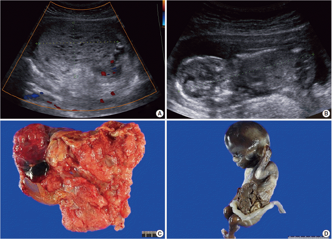
Fig. 1. Radiologic and gross findings. (A) Transabdominal ultrasound at 14+5 weeks of gestation shows a large multicystic honeycomb-like placenta measuring 8.8×5.7 cm with no sign of blood flow within the lesion. (B) The fetus has a crown-rump length of 9.6 cm. (C) The placenta is markedly enlarged with multiple vesicles. (D) The fetus exhibits gastroschisis.
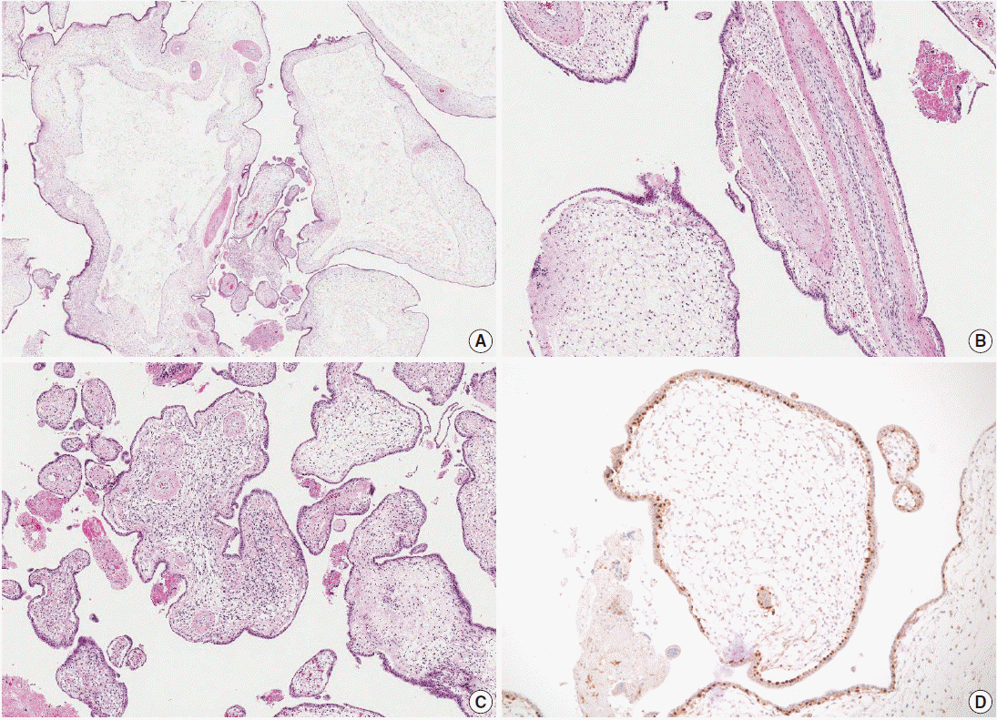
Fig. 2. Microscopic findings of the placenta. (A) Cystic vesicles are surrounded by stromal tissue and trophoblasts, indicating cysts in enlarged stem villi. No abnormal trophoblast proliferation is observed. (B) The majority of the enlarged stem villi have abnormal vessels with thickened muscular walls. (C) Some of the villi show mesenchymal cell proliferation. (D) Cytotrophoblasts are diffusely immunopositive for p57KIP2.
Reference
-
1. Vaisbuch E, Romero R, Kusanovic JP, et al. Three-dimensional sonography of placental mesenchymal dysplasia and its differential diagnosis. J Ultrasound Med. 2009; 28:359–68.
Article2. Moscoso G, Jauniaux E, Hustin J. Placental vascular anomaly with diffuse mesenchymal stem villous hyperplasia: a new clinico-pathological entity? Pathol Res Pract. 1991; 187:324–8.3. Arizawa M, Nakayama M. Suspected involvement of the X chromosome in placental mesenchymal dysplasia. Congenit Anom (Kyoto). 2002; 42:309–17.
Article4. Parveen Z, Tongson-Ignacio JE, Fraser CR, Killeen JL, Thompson KS. Placental mesenchymal dysplasia. Arch Pathol Lab Med. 2007; 131:131–7.
Article5. Pham T, Steele J, Stayboldt C, Chan L, Benirschke K. Placental mesenchymal dysplasia is associated with high rates of intrauterine growth restriction and fetal demise: a report of 11 new cases and a review of the literature. Am J Clin Pathol. 2006; 126:67–78.6. Heazell AE, Sahasrabudhe N, Grossmith AK, Martindale EA, Bhatia K. A case of intrauterine growth restriction in association with placental mesenchymal dysplasia with abnormal placental lymphatic development. Placenta. 2009; 30:654–7.
Article7. Woo GW, Rocha FG, Gaspar-Oishi M, Bartholomew ML, Thompson KS. Placental mesenchymal dysplasia. Am J Obstet Gynecol. 2011; 205:e3–5.
Article8. Ulker V, Aslan H, Gedikbasi A, Yararbas K, Yildirim G, Yavuz E. Placental mesenchymal dysplasia: a rare clinicopathologic entity confused with molar pregnancy. J Obstet Gynaecol. 2013; 33:246–9.
Article9. Feldkamp ML, Carey JC, Sadler TW. Development of gastroschisis: review of hypotheses, a novel hypothesis, and implications for research. Am J Med Genet A. 2007; 143A:639–52.
Article
- Full Text Links
-
- Actions
-
Cited
- CITED
-
- Close
- Share
-
- Similar articles
-
- Placental Mesenchymal Dysplasia Associated with Placenta Previa and Preterm Labor
- Placental Mesenchymal Dysplasia Associated with a Fetal Unilateral Multicystic Dysplastic Kidney: A Case Report
- Fetal Surgery: Gastroschisis Model in Rabbits
- Placental mesenchymal dysplasia associated with severe preeclampsia: A case report
- A case of gastroschisis associated with fetal death in utero, and ultrasonographic findings which were in antenatal period
