The Contribution of Resident Vascular Stem Cells to Arterial Pathology
- Affiliations
-
- 1Anatomic Pathology Institute, Department of Biomedicine and Prevention, Tor Vergata University of Rome, Rome, Italy. orlandi@uniroma2.it
- KMID: 2380794
- DOI: http://doi.org/10.15283/ijsc.2015.8.1.9
Abstract
- Intimal accumulation of smooth muscle cells contributes to the development and progression of atherosclerotic lesions and restenosis following endovascular procedures. Arterial smooth muscle cells display heterogeneous phenotypes in both physiological and pathological conditions. In response to injury, dedifferentiated or synthetic smooth muscle cells proliferate and migrate from the tunica media into the intima. As a consequence, smooth muscle cells in vascular lesions show a prevalent dedifferentiated phenotype compared to the contractile appearance of normal media smooth muscle cells. The discovery of abundant stem antigen-expressing cells in vascular lesions also rarely detected in the tunica media of normal adult vessels stimulated a great scientific debate concerning the possibility that proliferating vascular wall-resident stem cells accumulate into the neointima and contribute to the progression of lesions. Although several experimental studies support this hypothesis, others researchers suggest a positive effect of stem cells on plaque stabilization. So, the real contribute of vascular wall-resident stem cells to pathological vascular remodelling needs further investigation. This review will examine the evidence and the contribution of vascular wall-resident stem cells to arterial pathobiology, in order to address future investigations as potential therapeutic target to prevent the progression of vascular diseases.
Keyword
MeSH Terms
Figure
-

Fig. 1 phenotypic heterogeneity of adult vascular smooth muscle cells. Rat aortic normal media SMCs (left column) display with the classical “hill-and-valley” confluent grow pattern when cultured in plastic dishes, a more dendritic shape with a marked extracellular matrix remodelling when cultured in collagen gel and display abundant α-smooth muscle actin (α-sm actin)-positive stress fibers in immunofluorescence (rhodamine, bottom). In contrast, neointimal VSMCs obtained fifteen days after ballooning (right column) display a monolayered and epithelioid appearance (top), grow in Indian files with bipolar conjunctions in collagen gel and contain very low amount of α-smooth muscle actin (bottom).

Fig. 2 Stem cell expression in rat aorta after injury and with aging. Serial immunostainings reveal (A) very rare cell are c-kit+ positive in normal 2 month-old rat aorta. (B) fifteen days after ballon injury, the majority of intimal and some of medial SMCs are c-kit+ positive. (C) Very rare SMCs are flt-1+ in normal 2 month-old rat aorta, but (D) the number of flt-1+ cells increases in 24-month old rat aorta.
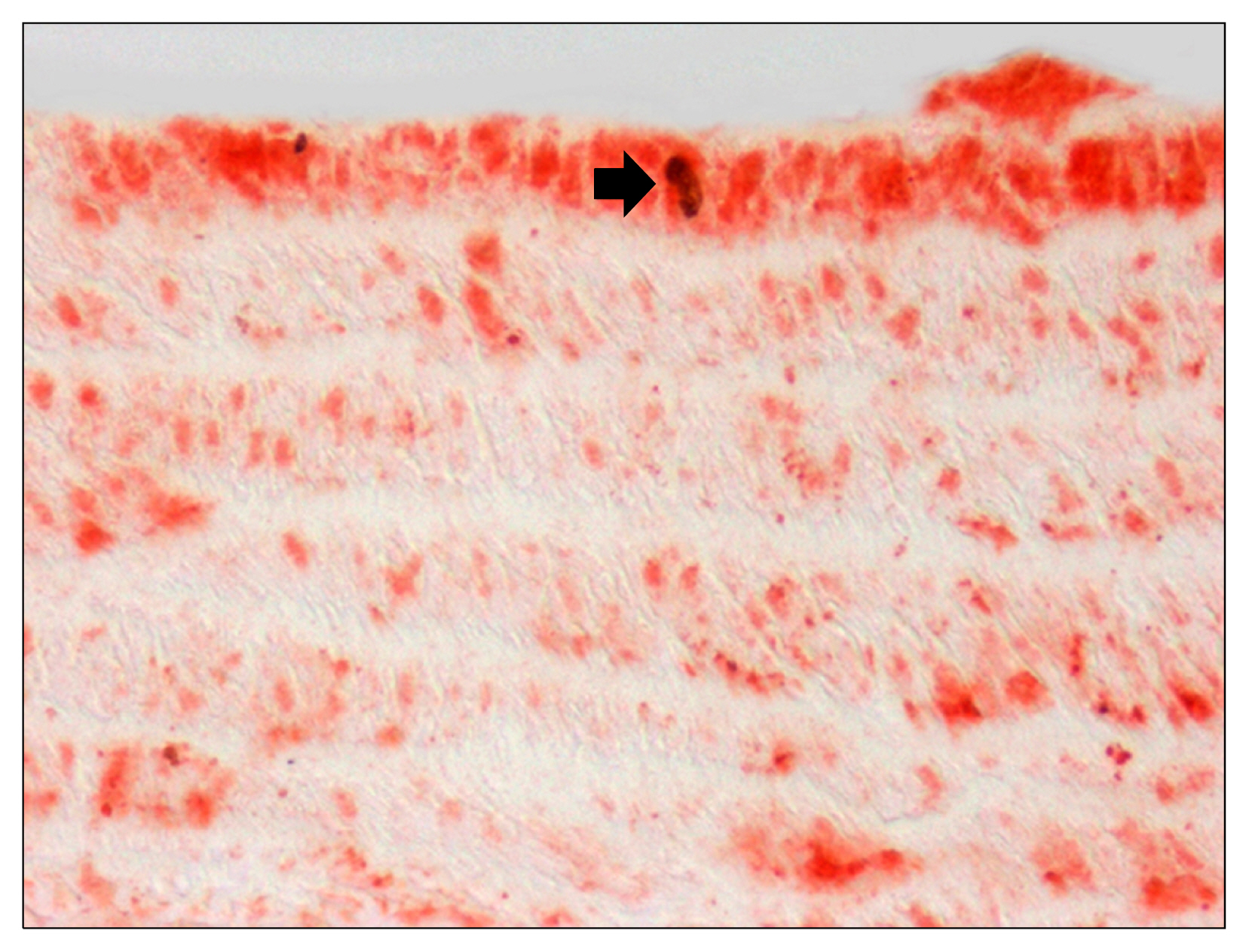
Fig. 3 flt-1 expression and smooth muscle cell proliferation. Double immunostaining reveals that flt-1 expression (red) characterizes rat aortic bromodeoxyurinine+ proliferating SMC (arrow, black) in the tunica media three days after ballooning.
Reference
-
References
1. Ross R, Glomset JA. The pathogenesis of atherosclerosis. Part 1. New Eng J Med. 1976; 295:369–377. DOI: 10.1056/NEJM197608122950707.2. Ross R, Glomset J. The pathogenesis of atherosclerosis (second of two parts). New Eng J Med. 1976; 295:420–425. DOI: 10.1056/NEJM197608192950805. PMID: 778621.3. Clowes AW, Reidy MA, Clowes MM. Kinetics of cellular proliferation after arterial injury. I. Smooth muscle growth in the absence of endothelium. Lab Invest. 1983; 49:327–333. PMID: 6887785.4. Gabbiani G. The cytoskeleton of rat aortic smooth muscle cells. Normal conditions, experimental intimal thickening, and tissue culture. Ann N Y Acad Sci. 1986; 488:196–198. DOI: 10.1111/j.1749-6632.1986.tb46558.x. PMID: 3472483.5. Benditt EP, Benditt JM. Evidence for a monoclonal origin of human atherosclerotic plaques. Proc Natl Acad Sci U S A. 1973; 70:1753–1756. DOI: 10.1073/pnas.70.6.1753. PMID: 4515934. PMCID: 433588.
Article6. Hu Y, Davison F, Ludewig B, Erdel M, Mayr M, Url M, Dietrich H, Xu Q. Smooth muscle cells in transplant atherosclerotic lesions are originated from recipients, but not bone marrow progenitor cells. Circulation. 2002; 106:1834–1839. DOI: 10.1161/01.CIR.0000031333.86845.DD. PMID: 12356638.
Article7. Sata M, Saiura A, Kunisato A, Tojo A, Okada S, Tokuhisa T, Hirai H, Makuuchi M, Hirata Y, Nagai R. Hematopoietic stem cells differentiate into vascular cells that participate in the pathogenesis of atherosclerosis. Nat Med. 2002; 8:403–409. DOI: 10.1038/nm0402-403. PMID: 11927948.
Article8. Caplice NM, Bunch TJ, Stalboerger PG, Wang S, Simper D, Miller DV, Russell SJ, Litzow MR, Edwards WD. Smooth muscle cells in human coronary atherosclerosis can originate from cells administered at marrow transplantation. Proc Natl Acad Sci U S A. 2003; 100:4754–4759. DOI: 10.1073/pnas.0730743100. PMID: 12665618. PMCID: 153628.
Article9. Bentzon JF, Weile C, Sondergaard CS, Hindkjaer J, Kassem M, Falk E. Smooth muscle cells in atherosclerosis originate from the local vessel wall and not circulating progenitor cells in ApoE knockout mice. Arterioscler Thromb Vasc Biol. 2006; 26:2696–2702. DOI: 10.1161/01.ATV.0000247243.48542.9d. PMID: 17008593.
Article10. Bentzon JF, Sondergaard CS, Kassem M, Falk E. Smooth muscle cells healing atherosclerotic plaque disruptions are of local, not blood, origin in apolipoprotein E knockout mice. Circulation. 2007; 116:2053–2061. DOI: 10.1161/CIRCULATIONAHA.107.722355. PMID: 17938286.
Article11. Torsney E, Mandal K, Halliday A, Jahangiri M, Xu Q. Characterisation of progenitor cells in human atherosclerotic vessels. Atherosclerosis. 2007; 191:259–264. DOI: 10.1016/j.atherosclerosis.2006.05.033.
Article12. Orlandi A, Di Lascio A, Francesconi A, Scioli MG, Arcuri G, Ferlosio A, Spagnoli LG. Stem cell marker expression and proliferation and apoptosis of vascular smooth muscle cells. Cell Cycle. 2008; 7:3889–3897. DOI: 10.4161/cc.7.24.7323. PMID: 19098424.
Article13. Gabbiani G, Kocher O, Bloom WS, Vandekerckhove J, Weber K. Actin expression in smooth muscle cells of rat aortic intimal thickening, human atheromatous plaque, and cultured rat aortic media. J Clin Invest. 1984; 73:148–152. DOI: 10.1172/JCI111185. PMID: 6690475. PMCID: 424985.
Article14. Orlandi A, Ehrlich HP, Ropraz P, Spagnoli LG, Gabbiani G. Rat aortic smooth muscle cells isolated from different layers and at different times after endothelial denudation show distinct biological features in vitro. Arterioscler Thromb. 1994; 14:982–989. DOI: 10.1161/01.ATV.14.6.982. PMID: 8199190.
Article15. Bochaton-Piallat ML, Ropraz P, Gabbiani F, Gabbiani G. Phenotypic heterogeneity of rat arterial smooth muscle cell clones. Implications for the development of experimental intimal thickening. Arterioscler Thromb Vasc Biol. 1996; 16:815–820. DOI: 10.1161/01.ATV.16.6.815. PMID: 8640410.16. Hao H, Ropraz P, Verin V, Camenzind E, Geinoz A, Pepper MS, Gabbiani G, Bochaton-Piallat ML. Heterogeneity of smooth muscle cell populations cultured from pig coronary artery. Arterioscler Thromb Vasc Biol. 2002; 22:1093–1099. DOI: 10.1161/01.ATV.0000022407.91111.E4. PMID: 12117722.
Article17. Li S, Sims S, Jiao Y, Chow LH, Pickering JG. Evidence from a novel human cell clone that adult vascular smooth muscle cells can convert reversibly between noncontractile and contractile phenotypes. Circ Res. 1999; 85:338–348. DOI: 10.1161/01.RES.85.4.338. PMID: 10455062.
Article18. Orlandi A, Ferlosio A, Gabbiani G, Spagnoli LG, Ehrlich PH. Phenotypic heterogeneity influences the behavior of rat aortic smooth muscle cells in collagen lattice. Exp Cell Res. 2005; 311:317–327. DOI: 10.1016/j.yexcr.2005.10.008. PMID: 16263112.
Article19. Orlandi A, Ropraz P, Gabbiani G. Proliferative activity and alpha-smooth muscle actin expression in cultured rat aortic smooth muscle cells are differently modulated by transforming growth factor-beta 1 and heparin. Exp Cell Res. 1994; 214:528–536. DOI: 10.1006/excr.1994.1290. PMID: 7925646.
Article20. Orlandi A, Francesconi A, Cocchia D, Corsini A, Spagnoli LG. Phenotypic heterogeneity influences apoptotic susceptibility to retinoic acid and cis-platinum of rat arterial smooth muscle cells in vitro: Implications for the evolution of experimental intimal thickening. Arterioscler Thromb Vasc Biol. 2001; 21:1118–1123. DOI: 10.1161/hq0701.092144. PMID: 11451739.
Article21. Orlandi A, Francesconi A, Marcellini M, Di Lascio A, Spagnoli LG. Propionyl-L-carnitine reduces proliferation and potentiates Bax-related apoptosis of aortic intimal smooth muscle cells by modulating nuclear factor-kappaB activity. J Biol Chem. 2007; 282:4932–4942.
Article22. Orlandi A, Ferlosio A, Arcuri G, Scioli MG, De Falco S, Spagnoli LG. Flt-1 expression influences apoptotic susceptibility of vascular smooth muscle cells through the NF-kappaB/IAP-1 pathway. Cardiovasc Res. 2010; 85:214–223.
Article23. Gittenberger-de Groot AC, DeRuiter MC, Bergwerff M, Poelmann RE. Smooth muscle cell origin and its relation to heterogeneity in development and disease. Arterioscler Thromb Vasc Biol. 1999; 19:1589–1594. PMID: 10397674.
Article24. Topouzis S, Majesky MW. Smooth muscle lineage diversity in the chick embryo. Two types of aortic smooth muscle cell differ in growth and receptor-mediated transcriptional responses to transforming growth factor-beta. Dev Biol. 1996; 178:430–445. PMID: 8830742.25. Suzuki S, Narita Y, Yamawaki A, Murase Y, Satake M, Mutsuga M, Okamoto H, Kagami H, Ueda M, Ueda Y. Effects of extracellular matrix on differentiation of human bone marrow-derived mesenchymal stem cells into smooth muscle cell lineage: utility for cardiovascular tissue engineering. Cells Tissues Organs. 2010; 191:269–280. DOI: 10.1159/000260061.
Article26. Simper D, Stalboerger PG, Panetta CJ, Wang S, Caplice NM. Smooth muscle progenitor cells in human blood. Circulation. 2002; 106:1199–1204. DOI: 10.1161/01.CIR.0000031525.61826.A8. PMID: 12208793.
Article27. Jiang Y, Jahagirdar BN, Reinhardt RL, Schwartz RE, Keene CD, Ortiz-Gonzalez XR, Reyes M, Lenvik T, Lund T, Blackstad M, Du J, Aldrich S, Lisberg A, Low WC, Largaespada DA, Verfaillie CM. Pluripotency of mesen-chymal stem cells derived from adult marrow. Nature. 2002; 418:41–49. DOI: 10.1038/nature00870. PMID: 12077603.
Article28. Sainz J, Al Haj Zen A, Caligiuri G, Demerens C, Urbain D, Lemitre M, Lafont A. Isolation of “side population” progenitor cells from healthy arteries of adult mice. Arterioscler Thromb Vasc Biol. 2006; 26:281–286.
Article29. Jackson KA, Mi T, Goodell MA. Hematopoietic potential of stem cells isolated from murine skeletal muscle. Proc Natl Acad Sci U S A. 1999; 96:14482–14486. PMID: 10588731. PMCID: 24462.
Article30. Tintut Y, Alfonso Z, Saini T, Radcliff K, Watson K, Boström K, Demer LL. Multilineage potential of cells from the artery wall. Circulation. 2003; 108:2505–2510. DOI: 10.1161/01.CIR.0000096485.64373.C5. PMID: 14581408.
Article31. Hu Y, Zhang Z, Torsney E, Afzal AR, Davison F, Metzler B, Xu Q. Abundant progenitor cells in the adventitia contribute to atherosclerosis of vein grafts in ApoE-deficient mice. J Clin Invest. 2004; 113:1258–1265. DOI: 10.1172/JCI19628. PMID: 15124016. PMCID: 398426.
Article32. Howson KM, Aplin AC, Gelati M, Alessandri G, Parati EA, Nicosia RF. The postnatal rat aorta contains pericyte progenitor cells that form spheroidal colonies in suspension culture. Am J Physiol Cell Physiol. 2005; 289:C1396–C13407. DOI: 10.1152/ajpcell.00168.2005. PMID: 16079185.
Article33. Hibbert B, Chen YX, O’Brien ER. c-kit-immunopositive vascular progenitor cells populate human coronary in-stent restenosis but not primary atherosclerotic lesions. Am J Physiol Heart Circ Physiol. 2004; 287:H518–H524. DOI: 10.1152/ajpheart.00002.2004. PMID: 15277195.
Article34. Spagnoli LG, Orlandi A, Santeusanio G. Foam cells of the rabbit atherosclerotic plaque arrested in metaphase by colchicine show a macrophage phenotype. Atherosclerosis. 1991; 88:87–92. DOI: 10.1016/0021-9150(91)90260-A. PMID: 1878013.
Article35. Spagnoli LG, Orlandi A, Marino B, Mauriello A, De Angelis C, Ramacci MT. Propionyl-L-carnitine prevents the progression of atherosclerotic lesions in aged hyperlipemic rabbits. Atherosclerosis. 1995; 114:29–44. DOI: 10.1016/0021-9150(94)05460-Z. PMID: 7605374.
Article36. Morrow D, Cullen JP, Liu W, Guha S, Sweeney C, Birney YA, Collins N, Walls D, Redmond EM, Cahill PA. Sonic Hedgehog induces Notch target gene expression in vascular smooth muscle cells via VEGF-A. Arterioscler Thromb Vasc Biol. 2009; 29:1112–1118. DOI: 10.1161/ATVBAHA.109.186890. PMID: 19407245. PMCID: 2794048.
Article37. Shi Y, Pieniek M, Fard A, O’Brien J, Mannion JD, Zalewski A. Adventitial remodeling after coronary arterial injury. Circulation. 1996; 93:340–348. DOI: 10.1161/01.CIR.93.2.340. PMID: 8548908.
Article38. Torsney E, Hu Y, Xu Q. Adventitial progenitor cells contribute to arteriosclerosis. Trends Cardiovasc Med. 2005; 15:64–68. DOI: 10.1016/j.tcm.2005.02.003. PMID: 15885572.
Article39. Hu Y, Zhang Z, Torsney E, Afzal AR, Davison F, Metzler B, Xu Q. Abundant progenitor cells in the adventitia contribute to atherosclerosis of vein grafts in ApoE-deficient mice. J Clin Invest. 2004; 113:1258–1265. DOI: 10.1172/JCI19628. PMID: 15124016. PMCID: 398426.
Article40. Gimble JM, Katz AJ, Bunnell BA. Adipose-derived stem cells for regenerative medicine. Circ Res. 2007; 100:1249–1260. DOI: 10.1161/01.RES.0000265074.83288.09. PMID: 17495232.
Article41. Orlandi A, Francesconi A, Marcellini M, Ferlosio A, Spagnoli LG. Role of ageing and coronary atherosclerosis in the development of cardiac fibrosis in the rabbit. Cardiovasc Res. 2004; 64:544–552. PMID: 15537508.
Article42. Cervelli V, Gentile P, Scioli MG, Grimaldi M, Casciani CU, Spagnoli LG, Orlandi A. Application of platelet-rich plasma in plastic surgery: clinical and in vitro evaluation. Tissue Eng Part C Methods. 2009; 15:625–634. PMID: 19231923.
Article43. Gentile P, Orlandi A, Scioli MG, Di Pasquali C, Bocchini I, Curcio CB, Floris M, Fiaschetti V, Floris R, Cervell V. A comparative translational study: the combined use of enhanced stromal vascular fraction and platelet-rich plasma improves fat grafting maintenance in breast reconstruction. Stem Cells Transl Med. 2012; 1:341–351. PMID: 23197813. PMCID: 3659694.
Article44. Cervelli V, Scioli MG, Gentile P, Doldo E, Bonanno E, Spagnoli LG, Orlandi A. Platelet-rich plasma greatly potentiates insulin-induced adipogenic differentiation of human adipose-derived stem cells through a serine/threonine kinase Akt-dependent mechanism and promotes clinical fat graft maintenance. Stem Cells Transl Med. 2012; 1:206–220. PMID: 23197780. PMCID: 3659852.
Article45. Scioli MG, Cervelli V, Arcuri G, Gentile P, Doldo E, Bielli A, Bonanno E, Orlandi A. High insulin-induced down-regulation of Erk-1/IGF-1R/FGFR-1 signaling is required for oxidative stress-mediated apoptosis of adipose-derived stem cells. J Cell Physiol. 2014; 229:2077–2087. PMID: 24818995.
Article46. Scioli MG, Bielli A, Gentile P, Mazzaglia D, Cervelli V, Orlandi A. The biomolecular basis of adipogenic differentiation of adipose-derived stem cells. Int J Mol Sci. 2014; 15:6517–6526. PMID: 24743893. PMCID: 4013644.
Article47. Cervelli V, Gentile P, De Angelis B, Calabrese C, Di Stefani A, Scioli MG, Curcio BC, Felici M, Orlandi A. Application of enhanced stromal vascular fraction and fat grafting mixed with PRP in post-traumatic lower extremity ulcers. Stem Cell Res. 2011; 6:103–111. PMID: 21195687.
Article48. Gentile P, Orlandi A, Scioli MG, Di Pasquali C, Bocchini I, Cervelli V. Concise review: adipose-derived stromal vascular fraction cells and platelet-rich plasma: basic and clinical implications for tissue engineering therapies in regenerative surgery. Stem Cells Transl Med. 2012; 1:230–236. PMID: 23197782. PMCID: 3659840.
Article49. Cervelli V, Garcovich S, Bielli A, Cervelli G, Curcio BC, Scioli MG, Orlandi A, Gentile P. The effect of autologous activated platelet rich plasma (AA-PRP) injection on pattern hair loss: clinical and histomorphometric evaluation. Biomed Res Int. 2014; 2014:760709. PMID: 24883322. PMCID: 4032742.
Article50. Cervelli V, Bocchini I, Di Pasquali C, De Angelis B, Cervelli G, Curcio CB, Orlandi A, Scioli MG, Tati E, Delogu P, Gentile P. P.R.L. platelet rich lipotransfert: our experience and current state of art in the combined use of fat and PRP. Biomed Res Int. 2013; 2013:434191. PMID: 24191244. PMCID: 3804297.
Article51. Bielli A, Scioli MG, Gentile P, Agostinelli S, Tarquini C, Cervelli V, Orlandi A. Adult adipose-derived stem cells and breast cancer: a controversial relationship. Springerplus. 2014; 3:345. DOI: 10.1186/2193-1801-3-345. PMID: 25089245. PMCID: 4117859.
Article52. De Angelis B, Gentile P, Orlandi F, Bocchini I, Di Pasquali C, Agovino A, Gizzi C, Patrizi F, Scioli MG, Orlandi A, Cervelli V. Limb Rescue: A New Autologous-Peripheral Blood Mononuclear Cells Technology in Critical Limb Ischemia and Chronic Ulcers. Tissue Eng Part C Methods. 2015; Epub ahead of print. DOI: 10.1089/ten.tec.2014.0245.
Article53. Koh YJ, Kang S, Lee HJ, Choi TS, Lee HS, Cho CH, Koh GY. Bone marrow-derived circulating progenitor cells fail to transdifferentiate into adipocytes in adult adipose tissues in mice. J Clin Invest. 2007; 117:3684–3695. DOI: 10.1172/JCI32504. PMID: 18060029. PMCID: 2096460.
Article54. Merfeld-Clauss S, Gollahalli N, March KL, Traktuev DO. Adipose tissue progenitor cells directly interact with endothelial cells to induce vascular network formation. Tissue Eng Part A. 2010; 16:2953–2966. DOI: 10.1089/ten.tea.2009.0635. PMID: 20486792. PMCID: 2928048.
Article55. Strassburg S, Nienhueser H, Stark GB, Finkenzeller G, Torio-Padron N. Human adipose-derived stem cells enhance the angiogenic potential of endothelial progenitor cells, but not of human umbilical vein endothelial cells. Tissue Eng Part A. 2013; 19:166–174. DOI: 10.1089/ten.tea.2011.0699.
Article56. Crisan M, Yap S, Casteilla L, Chen CW, Corselli M, Park TS, Andriolo G, Sun B, Zheng B, Zhang L, Norotte C, Teng PN, Traas J, Schugar R, Deasy BM, Badylak S, Buhring HJ, Giacobino JP, Lazzari L, Huard J, Péault B. A peri-vascular origin for mesenchymal stem cells in multiple human organs. Cell Stem Cell. 2008; 3:301–313. DOI: 10.1016/j.stem.2008.07.003. PMID: 18786417.
Article57. Zoll J, Fontaine V, Gourdy P, Barateau V, Vilar J, Leroyer A, Lopes-Kam I, Mallat Z, Arnal JF, Henry P, Tobelem G, Tedgui A. Role of human smooth muscle cell progenitors in atherosclerotic plaque development and composition. Cardiovasc Res. 2008; 77:471–480. DOI: 10.1093/cvr/cvm034.
Article58. Orlandi A, Bennett M. Progenitor cell-derived smooth muscle cells in vascular disease. Biochem Pharmacol. 2010; 79:1706–1713. DOI: 10.1016/j.bcp.2010.01.027. PMID: 20117099.
Article59. Orlandi A, Mauriello A, De Angelis C, Ramacci MT, Spagnoli LG. Age-related differences in the distribution and occurrence of atherosclerotic aortic lesions in the hyperlipemic rabbit. Arch Gerontol Geriatr. 1992; 15( Suppl 1):295–302. DOI: 10.1016/S0167-4943(05)80029-9. PMID: 18647699.
Article60. Spagnoli LG, Orlandi A, Mauriello A, Santeusanio G, de Angelis C, Lucreziotti R, Ramacci MT. Aging and atherosclerosis in the rabbit. 1. Distribution, prevalence and morphology of atherosclerotic lesions. Atherosclerosis. 1991; 89:11–24. DOI: 10.1016/0021-9150(91)90003-L. PMID: 1772469.61. Orlandi A, Mauriello A, Marino B, Spagnoli LG. Age-related modifications of aorta and coronaries in the rabbit: a morphological and morphometrical assessment. Arch Gerontol Geriatr. 1993; 17:37–53. DOI: 10.1016/0167-4943(93)90016-B. PMID: 15374330.
Article62. Spagnoli LG, Orlandi A, Mauriello A, De Angelis C, Ramacci MT. Age-dependent increase of rabbit aortic atherosclerosis. A morphometric approach. Pathol Res Pract. 1992; 188:637–642. DOI: 10.1016/S0344-0338(11)80071-3. PMID: 1409103.
Article63. Orlandi A, Francesconi A, Marcellini M, Ferlosio A, Spagnoli LG. Role of ageing and coronary atherosclerosis in the development of cardiac fibrosis in the rabbit. Cardiovasc Res. 2004; 64:544–552. DOI: 10.1016/j.cardiores.2004.07.024. PMID: 15537508.
Article64. Orlandi A, Francesconi A, Ferlosio A, Di Lascio A, Marcellini M, Pisano C, Spagnoli LG. Propionyl-L-carnitine prevents age-related myocardial remodeling in the rabbit. J Cardiovasc Pharmacol. 2007; 50:168–175. DOI: 10.1097/FJC.0b013e31805d8ee9. PMID: 17703133.
Article65. Ferlosio A, Arcuri G, Doldo E, Scioli MG, De Falco S, Spagnoli LG, Orlandi A. Age-related increase of stem marker expression influences vascular smooth muscle cell properties. Atherosclerosis. 2012; 224:51–57. DOI: 10.1016/j.atherosclerosis.2012.07.016. PMID: 22857896.
Article66. Campagnolo L, Costanza G, Francesconi A, Arcuri G, Moscatelli I, Orlandi A. Sortilin expression is essential for pro-nerve growth factor-induced apoptosis of rat vascular smooth muscle cells. PLoS One. 2014; 9:e84969. DOI: 10.1371/journal.pone.0084969. PMID: 24404198. PMCID: 3880332.
Article67. Tarallo V, Vesci L, Capasso O, Esposito MT, Riccioni T, Pastore L, Orlandi A, Pisano C, De Falco S. A placental growth factor variant unable to recognize vascular endothelial growth factor (VEGF) receptor-1 inhibits VEGF-dependent tumor angiogenesis via heterodimerization. Cancer Res. 2010; 70:1804–1813. DOI: 10.1158/0008-5472.CAN-09-2609. PMID: 20145150.
Article68. Cassinelli G, Zuco V, Petrangolini G, De Cesare M, Tortoreto M, Lanzi C, Cominetti D, Zaffaroni N, Orlandi A, Passeri D, Meco D, Di Francesco AM, Riccardi R, Bucci F, Pisano C, Zunino F. The curative efficacy of namitecan (ST1968) in preclinical models of pediatric sarcoma is associated with antiangiogenic effects. Biochem Pharmacol. 2012; 84:163–171. DOI: 10.1016/j.bcp.2012.04.005. PMID: 22525722.
Article69. Orlandi A, Bochaton-Piallat ML, Gabbiani G, Spagnoli LG. Aging, smooth muscle cells and vascular pathobiology: implications for atherosclerosis. Atherosclerosis. 2006; 188:221–230. DOI: 10.1016/j.atherosclerosis.2006.01.018. PMID: 16487530.
Article70. Stasi MA, Scioli MG, Arcuri G, Mattera GG, Lombardo K, Marcellini M, Riccioni T, De Falco S, Pisano C, Spagnoli LG, Borsini F, Orlandi A. Propionyl-L-carnitine improves postischemic blood flow recovery and arteriogenetic revascularization and reduces endothelial NADPH-oxidase 4-mediated superoxide production. Arterioscler Thromb Vasc Biol. 2010; 30:426–435. DOI: 10.1161/ATVBAHA.109.201533. PMID: 20056909.
Article
- Full Text Links
-
- Actions
-
Cited
- CITED
-
- Close
- Share
-
- Similar articles
-
- Roles of Vascular Smooth Muscle Cells in Atherosclerotic Calcification
- Is Stem Cell-Based Therapy Going on or Out for Cardiac Disease?
- Endogenous Gastric-Resident Mesenchymal Stem Cells Contribute to Formation of Cancer Stroma and Progression of Gastric Cancer
- Vascularized Organoids: A More Complete Model
- The Role of Microenvironment in Preserving the Potency of Adult Porcine Pulmonary Valve Stem Cells In Vitro
