Eosinophilic Gastritis Presenting as Tissue Necrosis
- Affiliations
-
- 1Department of Internal Medicine, Dong-A University College of Medicine, Busan, Korea.
- 2Department of Pathology, Dong-A University College of Medicine, Busan, Korea. jsjang@dau.ac.kr
- KMID: 2380416
- DOI: http://doi.org/10.5946/ce.2015.48.6.558
Abstract
- Eosinophilic gastroenteritis is very rare disorder that is characterized by eosinophilic infiltration of the gastrointestinal tract in the absence of any definite causes of eosinophilia. It is associated with various clinical gastrointestinal manifestations, and depends on the involved layer and site. We report a case of eosinophilic gastritis presenting with severe necrosis. The symptoms disappeared immediately after beginning steroid treatment, and the eosinophil count decreased to the reference range. The patient showed eosinophilic gastritis characterized by necrotic change such as necrotizing gastritis. It is a unique presentation of eosinophilic gastritis. To the best of our knowledge, no case of eosinophilic gastritis characterized by necrotic change such as necrotizing gastritis has been previously reported in Korea.
Keyword
MeSH Terms
Figure
-
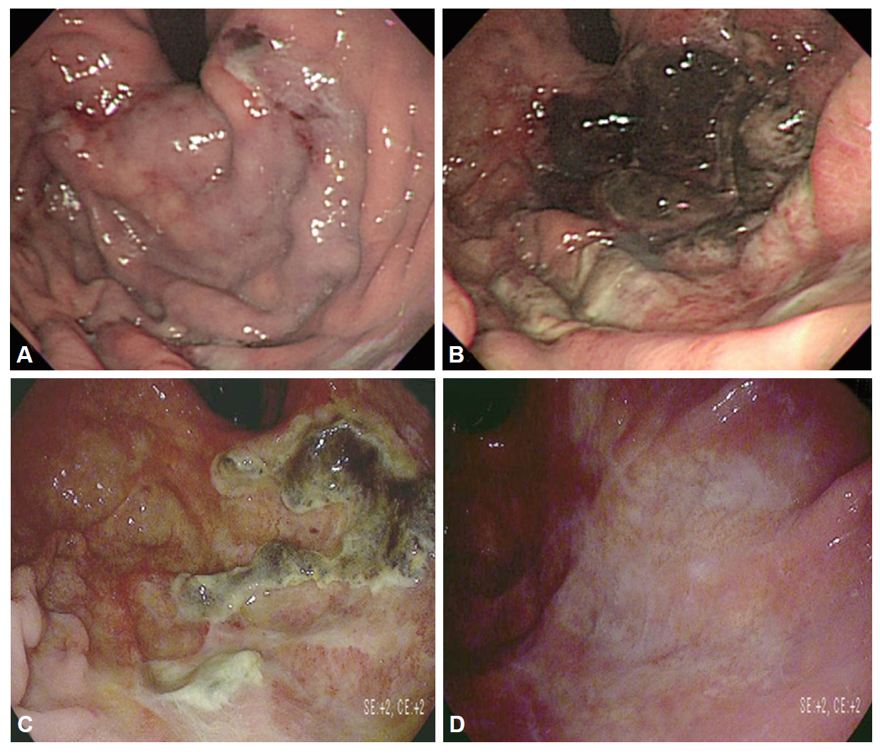
Fig. 1. (A) The first esophagogastroduodenoscopy (EGD) revealing multiple focal ulcerative lesions with diffuse discoloration and edematous change of the rugae in the gastric fundus, cardia, and upper body. (B) The second EGD showing diffuse necrotic change in the fundus, cardia, and upper body. (C) After 7 days of steroid treatment, the third EGD showing regenerative epithelial tissue with peeling off, of the necrotic tissue. (D) The fourth EGD showing the replacement of white scar tissue.
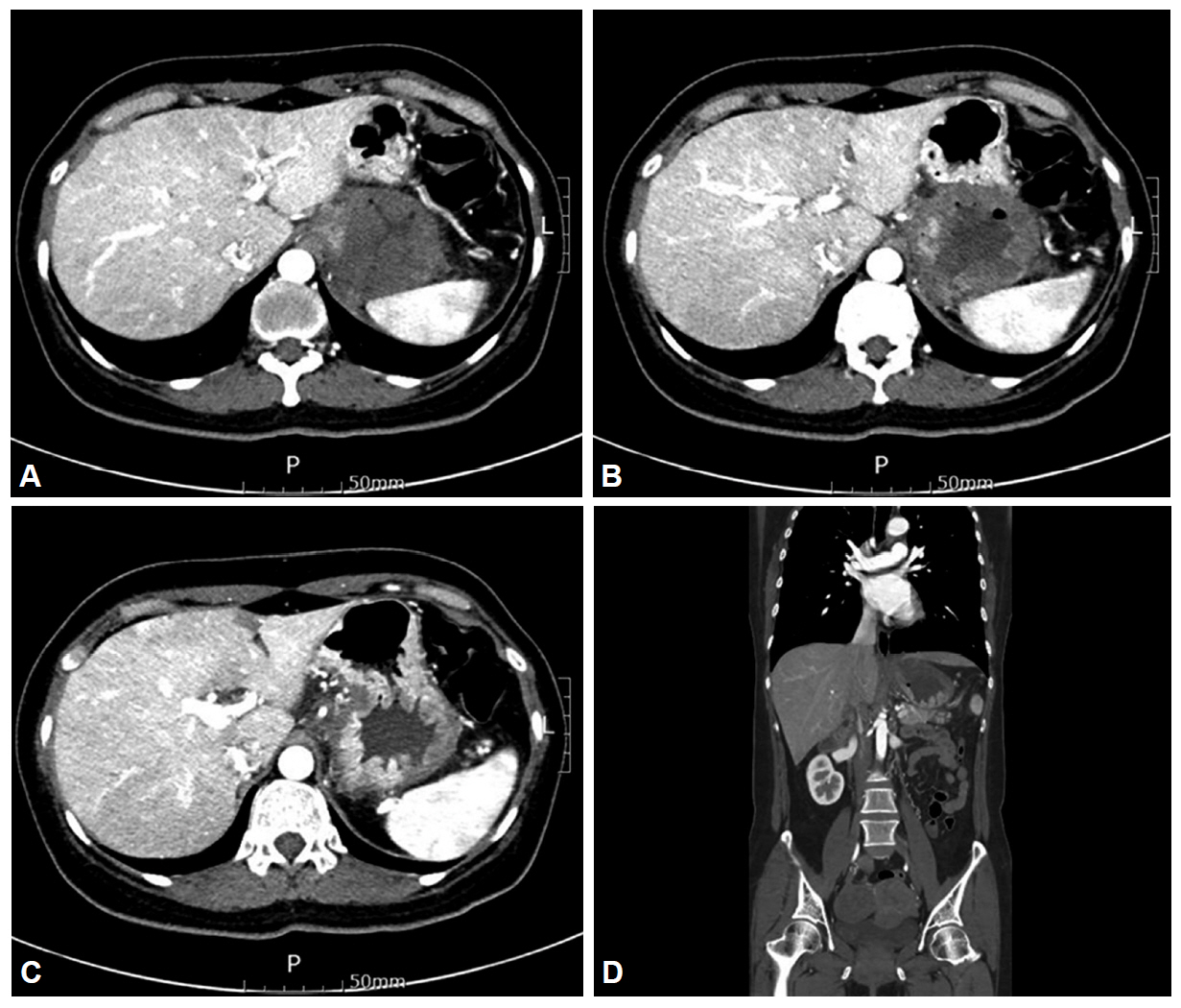
Fig. 2. (A-D) Abdominal computed tomography revealing severe edematous wall thickening with focal localized low attenuation of the fundus and cardia of the stomach.
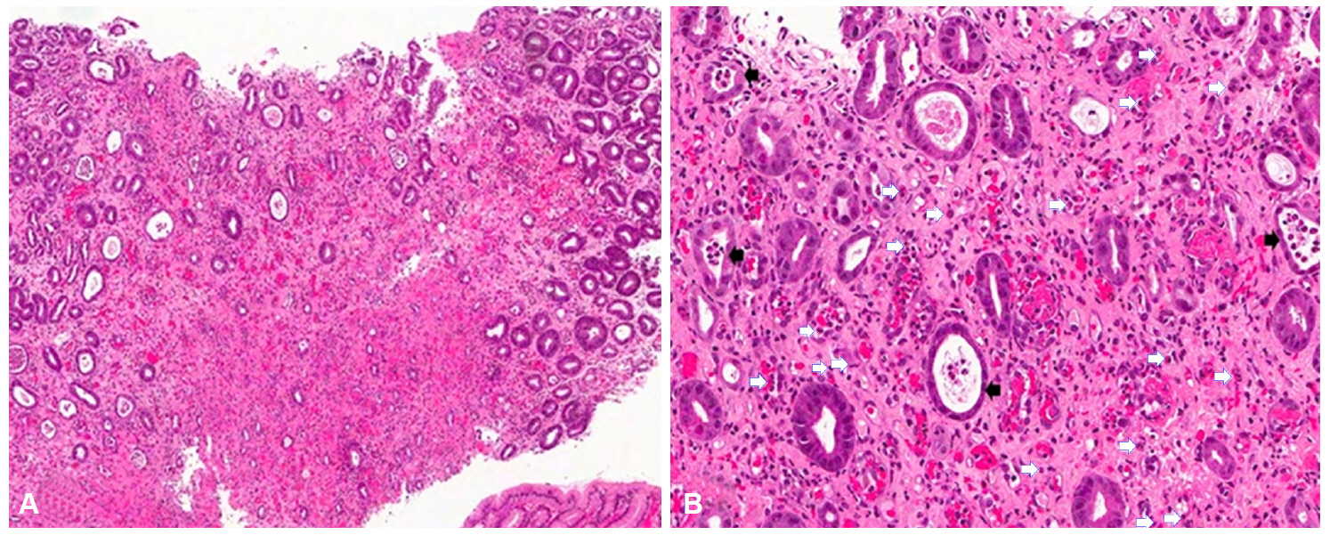
Fig. 3. Histological examination of gastric mucosal biopsy (H&E stain; A, ×100; B, ×200). The stomach showing erosion, dense infiltration of eosinophils in the lamina propria (white arrows), and glandular atrophy with crypt abscess (black arrows).
Reference
-
1. Rothenberg ME. Eosinophilic gastrointestinal disorders (EGID). J Allergy Clin Immunol. 2004; 113:11–28.
Article2. Talley NJ, Shorter RG, Phillips SF, Zinsmeister AR. Eosinophilic gastroenteritis: a clinicopathological study of patients with disease of the mucosa, muscle layer, and subserosal tissues. Gut. 1990; 31:54–58.
Article3. Yan BM, Shaffer EA. Primary eosinophilic disorders of the gastrointestinal tract. Gut. 2009; 58:721–732.
Article4. Navab F, Kleinman MS, Algazy K, Schenk E, Turner MD. Endoscopic diagnosis of eosinophilic gastritis. Gastrointest Endosc. 1972; 19:67–69.
Article5. Chowdhury A, Dhali GK, Banerjee PK. Etiology of gastric outlet obstruction. Am J Gastroenterol. 1996; 91:1679.6. Hogan SP, Rothenberg ME. Eosinophil function in eosinophil-associated gastrointestinal disorders. Curr Allergy Asthma Rep. 2006; 6:65–71.
Article7. Siewert E, Lammert F, Koppitz P, Schmidt T, Matern S. Eosinophilic gastroenteritis with severe protein-losing enteropathy: successful treatment with budesonide. Dig Liver Dis. 2006; 38:55–59.
Article8. Baig MA, Qadir A, Rasheed J. A review of eosinophilic gastroenteritis. J Natl Med Assoc. 2006; 98:1616–1619.9. Liacouras CA, Wenner WJ, Brown K, Ruchelli E. Primary eosinophilic esophagitis in children: successful treatment with oral corticosteroids. J Pediatr Gastroenterol Nutr. 1998; 26:380–385.
Article10. Daneshjoo R, N JT. Eosinophilic gastroenteritis. Curr Gastroenterol Rep. 2002; 4:366–372.
Article11. Neustrom MR, Friesen C. Treatment of eosinophilic gastroenteritis with montelukast. J Allergy Clin Immunol. 1999; 104(2 Pt 1):506.
Article12. Suzuki J, Kawasaki Y, Nozawa R, et al. Oral disodium cromoglycate and ketotifen for a patient with eosinophilic gastroenteritis, food allergy and protein-losing enteropathy. Asian Pac J Allergy Immunol. 2003; 21:193–197.13. Strauss RJ, Friedman M, Platt N, Gassner W, Wise L. Gangrene of the stomach: a case of acute necrotizing gastritis. Am J Surg. 1978; 135:253–257.
Article14. Pohl JF, Melin-Aldana H, Rudolph C. Prolapse gastropathy in the pediatric patient. J Pediatr Gastroenterol Nutr. 2000; 30:458–460.
Article15. Bishop PR, Nowicki MJ, Parker PH. Vomiting-induced hematemesis in children: Mallory-Weiss tear or prolapse gastropathy? J Pediatr Gastroenterol Nutr. 2000; 30:436–441.
Article
- Full Text Links
-
- Actions
-
Cited
- CITED
-
- Close
- Share
-
- Similar articles
-
- A Case of Eosinophilic Gastritis with Delayed Gastric Emptying
- Radiographic and pathologic observations of eosinophilic gastroenteritis
- Praziquantel Treatment of Eosinophilic Gastritis Suspected to Be Due to Cerebral Sparganosis
- A Case of Eosinophilic Gastroenteritis with Rapid Deterioration Mimicking Borrmann Type 4 Advanced Gastric Cancer
- A Case of Non-IgE-mediated Eosinophilic Gastroenteritis Presenting as Ascites
