Endoscopic Submucosal Dissection for Recurrent or Residual Superficial Esophageal Cancer after Chemoradiotherapy: Two Cases
- Affiliations
-
- 1Department of Internal Medicine, Gangnam Severance Hospital, Yonsei University College of Medicine, Seoul, Korea. DRYOUN@yuhs.ac
- 2Department of Diagnostic Pathology, Gangnam Severance Hospital, Yonsei University College of Medicine, Seoul, Korea.
- KMID: 2380415
- DOI: http://doi.org/10.5946/ce.2015.48.6.553
Abstract
- We report two cases of endoscopic submucosal dissection (ESD) for recurrent or residual esophageal squamous cell carcinoma (ESCC) lesions after chemoradiotherapy for advanced esophageal cancer. Case 1 involved a 64-year-old man who had previously undergone chemoradiotherapy for advanced ESCC and achieved a complete response (CR) for 22 months, until metachronous recurrent superficial ESCC was detected on follow-up esophagogastroduodenoscopy (EGD). We performed ESD and found no evidence of recurrence for 24 months. Case 2 involved a 59-year-old man who had previously undergone chemoradiotherapy for advanced ESCC. He responded favorably to treatment, and most of the tumor had disappeared on follow-up EGD 4 months later. However, there were two residual superficial esophageal lugol-voiding lesions. We performed ESD, and he had a CR for 32 months thereafter. ESD can be considered a viable treatment option for recurrent or residual superficial ESCC after chemoradiotherapy for advanced esophageal cancer.
MeSH Terms
Figure
-
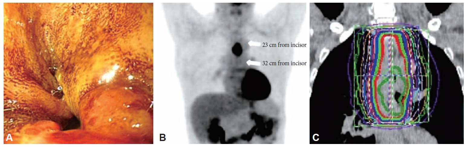
Fig. 1. Initial esophagogastroduodenoscopy and positron emission tomography (PET) findings of case 1. (A) Esophageal mass lesion 24 cm from the incisors causing malignant stricture; the scope could not reach past the lesion. (B) PET image showing esophageal cancer at the mid to distal portion with paraesophageal invasion, including invasion of the subcarinal lymph node. (C) Image showing the radiation field.
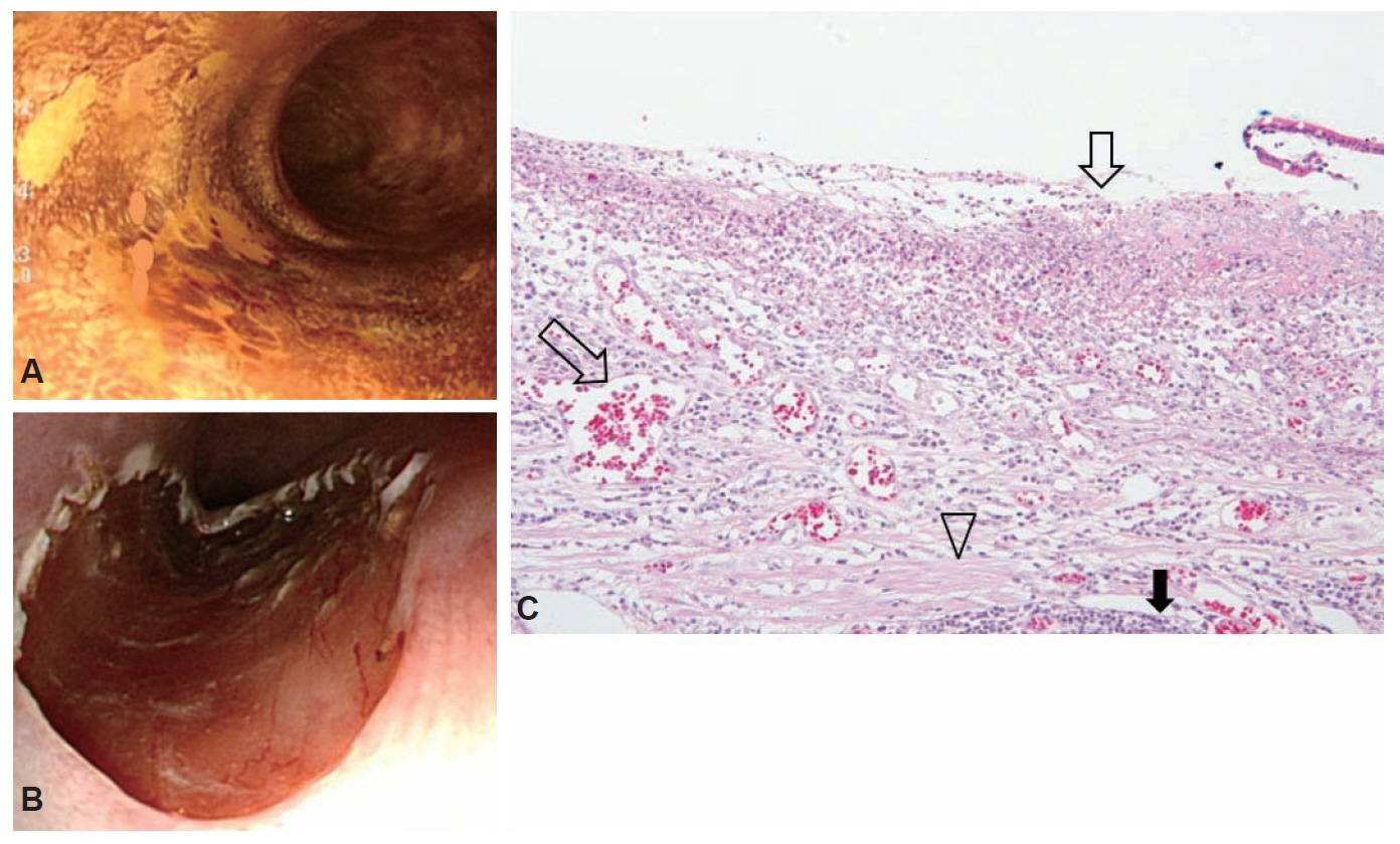
Fig. 2. Esophagogastroduodenoscopy procedure and pathologic findings of case 1. (A, B) Salvage endoscopic submucosal dissection (ESD) for metachronous recurrent superficial esophageal squamous cell carcinoma. The lesion is 32 cm from the incisors and previously treated with radiation. (C) The pathologic finding of the ESD specimen showing a well-differentiated squamous cell carcinoma with lamina propria invasion. Ulceration with dilated vessels (white arrows), inflammatory infiltrates (black arrow), and mild fibrosis (arrowhead) in the lamina propria, so-called radiation change, are noted (H&E stain, ×200).

Fig. 3. Initial esophagogastroduodenoscopy (EGD) and positron emission tomography (PET) findings of case 2. (A) EGD image showing an esophageal ulcerofungating mass. (B) PET scan showing a large esophageal lesion 29 to 43 cm from the incisors and metastasis to the left supraclavicular lymph node (white arrows). (C) Image showing the radiation field.

Fig. 4. Esophagogastroduodenoscopy procedure and pathologic finding of case 2. (A, B) Endoscopic submucosal dissection (ESD) en bloc resection of the residual superficial lugol-voiding lesion 38 cm from the incisors. (C) The pathologic finding of the ESD specimen showing a well-differentiated squamous cell carcinoma with lamina propria invasion. Dilated vessels (white arrow), inflammatory infiltrates (black arrow), and mild fibrosis (arrowhead) in the lamina propria are shown (H&E stain, ×200).
Reference
-
1. Meunier B, Raoul J, Le Prisé E, Lakéhal M, Launois B. Salvage esophagectomy after unsuccessful curative chemoradiotherapy for squamous cell cancer of the esophagus. Dig Surg. 1998; 15:224–226.
Article2. Swisher SG, Wynn P, Putnam JB, et al. Salvage esophagectomy for recurrent tumors after definitive chemotherapy and radiotherapy. J Thorac Cardiovasc Surg. 2002; 123:175–183.
Article3. Saito Y, Takisawa H, Suzuki H, et al. Endoscopic submucosal dissection of recurrent or residual superficial esophageal cancer after chemoradiotherapy. Gastrointest Endosc. 2008; 67:355–359.
Article4. Yano T, Muto M, Hattori S, et al. Long-term results of salvage endoscopic mucosal resection in patients with local failure after definitive chemoradiotherapy for esophageal squamous cell carcinoma. Endoscopy. 2008; 40:717–721.
Article5. Fujishiro M, Yahagi N, Kakushima N, et al. Endoscopic submucosal dissection of esophageal squamous cell neoplasms. Clin Gastroenterol Hepatol. 2006; 4:688–694.
Article6. Ishihara R, Iishi H, Uedo N, et al. Comparison of EMR and endoscopic submucosal dissection for en bloc resection of early esophageal cancers in Japan. Gastrointest Endosc. 2008; 68:1066–1072.
Article7. Stahl M, Stuschke M, Lehmann N, et al. Chemoradiation with and without surgery in patients with locally advanced squamous cell carcinoma of the esophagus. J Clin Oncol. 2005; 23:2310–2317.
Article8. Takeuchi M, Kobayashi M, Hashimoto S, et al. Salvage endoscopic submucosal dissection in patients with local failure after chemoradiotherapy for esophageal squamous cell carcinoma. Scand J Gastroenterol. 2013; 48:1095–1101.
Article9. Koizumi S, Jin M, Matsuhashi T, et al. Salvage endoscopic submucosal dissection for the esophagus-localized recurrence of esophageal squamous cell cancer after definitive chemoradiotherapy. Gastrointest Endosc. 2014; 79:348–353.
Article10. Mochizuki Y, Saito Y, Tanaka T, et al. Endoscopic submucosal dissection combined with the placement of biodegradable stents for recurrent esophageal cancer after chemoradiotherapy. J Gastrointest Cancer. 2012; 43:324–328.
Article
- Full Text Links
-
- Actions
-
Cited
- CITED
-
- Close
- Share
-
- Similar articles
-
- Endoscopic Submucosal Dissection Followed by Concurrent Chemoradiotherapy in Patients with Early Esophageal Cancer with a High Risk of Lymph Node Metastasis
- Endoscopic Treatment for Esophageal Cancer
- Photodynamic Therapy for Esophageal Cancer
- Salvage Endoscopic Resection for Residual Lesion after Definitive Chemoradiotherapy in Esophageal Cancer
- Superficial Esophageal Neoplasms Overlying Leiomyomas Removed by Endoscopic Submucosal Dissection: Case Reports and Review of the Literature
