Ultra-Low-Dose Chest CT in Patients with Neutropenic Fever and Hematologic Malignancy: Image Quality and Its Diagnostic Performance
- Affiliations
-
- 1Department of Radiology and Center for Imaging Science, Samsung Medical Center, Sungkyunkwan University School of Medicine, Seoul, Korea. hoyunlee96@gmail.com
- 2Division of Respiratory and Critical Care Medicine, Department of Internal Medicine, Samsung Medical Center, Sungkyunkwan University School of Medicine, Seoul, Korea.
- KMID: 2380377
- DOI: http://doi.org/10.4143/crt.2013.132
Abstract
- PURPOSE
The aim of this study was to evaluate the image quality of ultra-low-dose computed tomography (ULDCT) and its diagnostic performance in making a specific diagnosis of pneumonia in febrile neutropenic patients with hematological malignancy.
MATERIALS AND METHODS
ULDCT was performed prospectively in 207 febrile neutropenic patients with hematological malignancy. Three observers independently recorded the presence of lung parenchymal abnormality, and also indicated the cause of the lung parenchymal abnormality between infectious and noninfectious causes. If infectious pneumonia was considered the cause of lung abnormalities, they noted the two most appropriate diagnoses among four infectious conditions, including fungal, bacterial, viral, and Pneumocystis pneumonia. Sensitivity for correct diagnoses and receiver operating characteristic (ROC) curve analysis for evaluation of diagnostic accuracy were calculated. Interobserver agreements were determined using intraclass correlation coefficient.
RESULTS
Of 207 patients, 139 (67%) had pneumonia, 12 had noninfectious lung disease, and 56 had no remarkable chest computed tomography (CT) (20 with extrathoracic fever focus and 36 with no specific disease). Mean radiation expose dose of ULDCT was 0.60+/-0.15 mSv. Each observer regarded low-dose CT scans as unacceptable in only four (1.9%), one (0.5%), and three (1.5%) cases of ULDCTs. Sensitivity and area under the ROC curve in making a specific pneumonia diagnosis were 63.0%, 0.65 for reader 1; 63.0%, 0.61 for reader 2; and 65.0%, 0.62 for reader 3; respectively
CONCLUSION
ULDCT, with a sub-mSv radiation dose and acceptable image quality, provides ready and reasonably acceptable diagnostic information for pulmonary infection in febrile neutropenic patients with hematologic malignancy
MeSH Terms
Figure
-
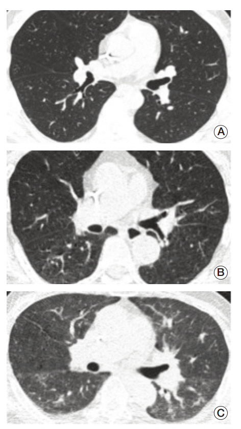
Fig. 1. Representative figures for assessment of image quality. (A) Excellent image quality. (B) Acceptable image quality. (C) Unacceptable image quality.

Fig. 2. Representative cases for infectious pneumonia. (A) Transverse ultra-low-dose computed tomography (ULDCT) scan in a patient with Aspergillus infection shows a nodule surrounded by a halo of ground-glass opacity in the left lower lobe (arrow). All three observers reached a consensus as an excellent image quality level. (B) Transverse ULDCT scans in a patient with Pneumocystis pneumonia show bilateral patchy areas of ground-glass opacity (arrows). All three observers reached a consensus as an acceptable image quality level. (C) Transverse and coronal ULDCT scans in a patient with streptococcal pneumonia show consolidation involving the posterior basal segment of the right lower lobe (arrows). All three observers reached a consensus as an acceptable image quality level. (D) Transverse ULDCT scans in a patient with coronavirus infection show multiple centrilobular nodules (arrowheads) and bilateral areas of lobular consolidation of peribronchial distributions (arrows). All three observers reached a consensus as an unacceptable image quality level.
Reference
-
References
1. de Naurois J, Novitzky-Basso I, Gill MJ, Marti FM, Cullen MH, Roila F, et al. Management of febrile neutropenia: ESMO Clinical Practice Guidelines. Ann Oncol. 2010; 21 Suppl 5:v252-6.
Article2. Maschmeyer G, Link H, Hiddemann W, Meyer P, Helmerking M, Eisenmann E, et al. Pulmonary infiltrations in febrile patients with neutropenia: risk factors and outcome under empirical antimicrobial therapy in a randomized multicenter study. Cancer. 1994; 73:2296–304.
Article3. Freifeld AG, Bow EJ, Sepkowitz KA, Boeckh MJ, Ito JI, Mullen CA, et al. Clinical practice guideline for the use of antimicrobial agents in neutropenic patients with cancer: 2010 update by the infectious diseases society of america. Clin Infect Dis. 2011; 52:e56–93.
Article4. Heussel CP, Kauczor HU, Heussel GE, Fischer B, Begrich M, Mildenberger P, et al. Pneumonia in febrile neutropenic patients and in bone marrow and blood stem-cell transplant recipients: use of high-resolution computed tomography. J Clin Oncol. 1999; 17:796–805.
Article5. Rizzi EB, Schinina V, Gentile FP, Bibbolino C. Reduced computed tomography radiation dose in HIV-related pneumonia: effect on diagnostic image quality. Clin Imaging. 2007; 31:178–84.
Article6. Stanzani M, Battista G, Sassi C, Lewis RE, Tolomelli G, Clissa C, et al. Computed tomographic pulmonary angiography for diagnosis of invasive mold diseases in patients with hematological malignancies. Clin Infect Dis. 2012; 54:610–6.
Article7. Brenner DJ, Hall EJ. Computed tomography: an increasing source of radiation exposure. N Engl J Med. 2007; 357:2277–84.8. Pearce MS, Salotti JA, Little MP, McHugh K, Lee C, Kim KP, et al. Radiation exposure from CT scans in childhood and subsequent risk of leukaemia and brain tumours: a retrospective cohort study. Lancet. 2012; 380:499–505.
Article9. Hendee WR, O'Connor MK. Radiation risks of medical imaging: separating fact from fantasy. Radiology. 2012; 264:312–21.
Article10. Kalra MK, Maher MM, Toth TL, Hamberg LM, Blake MA, Shepard JA, et al. Strategies for CT radiation dose optimization. Radiology. 2004; 230:619–28.
Article11. Patsios D, Maimon N, Chung T, Roberts H, Disperati P, Minden M, et al. Chest low-dose computed tomography in neutropenic acute myeloid leukaemia patients. Respir Med. 2010; 104:600–5.
Article12. Takahashi M, Maguire WM, Ashtari M, Khan A, Papp Z, Alberico R, et al. Low-dose spiral computed tomography of the thorax: comparison with the standard-dose technique. Invest Radiol. 1998; 33:68–73.13. Udayasankar UK, Li J, Baumgarten DA, Small WC, Kalra MK. Acute abdominal pain: value of non-contrast enhanced ultra-low-dose multi-detector row CT as a substitute for abdominal radiographs. Emerg Radiol. 2009; 16:61–70.
Article14. Caillot D, Couaillier JF, Bernard A, Casasnovas O, Denning DW, Mannone L, et al. Increasing volume and changing characteristics of invasive pulmonary aspergillosis on sequential thoracic computed tomography scans in patients with neutropenia. J Clin Oncol. 2001; 19:253–9.
Article15. Franquet T. Imaging of pulmonary viral pneumonia. Radiology. 2011; 260:18–39.
Article16. Heussel CP, Kauczor HU, Ullmann AJ. Pneumonia in neutropenic patients. Eur Radiol. 2004; 14:256–71.
Article17. Mayo JR, Aldrich J, Muller NL; Fleischner Society. Radiation exposure at chest CT: a statement of the Fleischner Society. Radiology. 2003; 228:15–21.
Article18. Rossini F, Verga M, Pioltelli P, Giltri G, Sancassani V, Pogliani EM, et al. Incidence and outcome of pneumonia in patients with acute leukemia receiving first induction therapy with anthracycline-containing regimens. Haematologica. 2000; 85:1255–60.19. Quadri TL, Brown AE. Infectious complications in the critically ill patient with cancer. Semin Oncol. 2000; 27:335–46.20. Donowitz GR, Harman C, Pope T, Stewart FM. The role of the chest roentgenogram in febrile neutropenic patients. Arch Intern Med. 1991; 151:701–4.
Article21. Korones DN, Hussong MR, Gullace MA. Routine chest radiography of children with cancer hospitalized for fever and neutropenia: is it really necessary? Cancer. 1997; 80:1160–4.22. Neofytos D. Chest computed tomography versus serum galactomannan enzyme immunoassay for the diagnosis of probable invasive aspergillosis: to be decided. Clin Infect Dis. 2010; 51:1281–3.
Article23. Prakash P, Kalra MK, Digumarthy SR, Hsieh J, Pien H, Singh S, et al. Radiation dose reduction with chest computed tomography using adaptive statistical iterative reconstruction technique: initial experience. J Comput Assist Tomogr. 2010; 34:40–5.24. Heussel CP, Kauczor HU, Heussel G, Fischer B, Mildenberger P, Thelen M. Early detection of pneumonia in febrile neutropenic patients: use of thin-section CT. AJR Am J Roentgenol. 1997; 169:1347–53.
Article25. Neroladaki A, Botsikas D, Boudabbous S, Becker CD, Montet X. Computed tomography of the chest with model-based iterative reconstruction using a radiation exposure similar to chest X-ray examination: preliminary observations. Eur Radiol. 2013; 23:360–6.
Article
- Full Text Links
-
- Actions
-
Cited
- CITED
-
- Close
- Share
-
- Similar articles
-
- Performance of Half-dose Chest Computed Tomography in Lung Malignancy Using an Iterative Reconstruction Technique
- Detection of Pulmonary Metastatic Nodules: Usefulness of Low-dose Multidetector CT in Comparison with Chest Radiograph
- Evaluation of Ultra-Low Dose CT in the Diagnosis of Pediatric-Like Fractures Using an Experimental Animal Study
- Urinary tract infections in pediatric oncology patients with febrile neutropenia
- Dose and Image Evaluations of Imaging for Radiotherapy
