A Case of Teratoma of Thyroid Gland in Adolescence
- Affiliations
-
- 1Division of Endocrinology and Metabolism, Department of Internal Medicine, Sejong General Hospital, Bucheon, Korea. tusyle@naver.com
- KMID: 2379364
- DOI: http://doi.org/10.11106/ijt.2017.10.1.61
Abstract
- Benign teratomas of the thyroid are very rare in adolescence and adults. In this review, we report a case of 14-year-old Korean girl with huge neck mass. She presented with anterior neck enlargement and compression symptom which was rapidly aggravated in 2 months. In physical examination, enlarged and firm right thyroid lobe was palpated and laboratory test of thyroid function was normal. In ultrasonography, right lobe was mainly occupied with a solid nodule size of 44×23×25 mm, showing markedly inhomogeneous hypoechogenicity. As fine needle aspiration cytology was non-diagnostic, lobectomy was done. Histological examination demonstrated that the tumor is benign thyroid teratoma composed of tissue from all three germ layers. When large thyroid nodule is detected in adolescence and malignancy could not be ruled out, final diagnosis should be made with surgical resection. And we should at least attentive for possibility of teratomas when ultrasonographic findings are suspicious.
Keyword
MeSH Terms
Figure
-
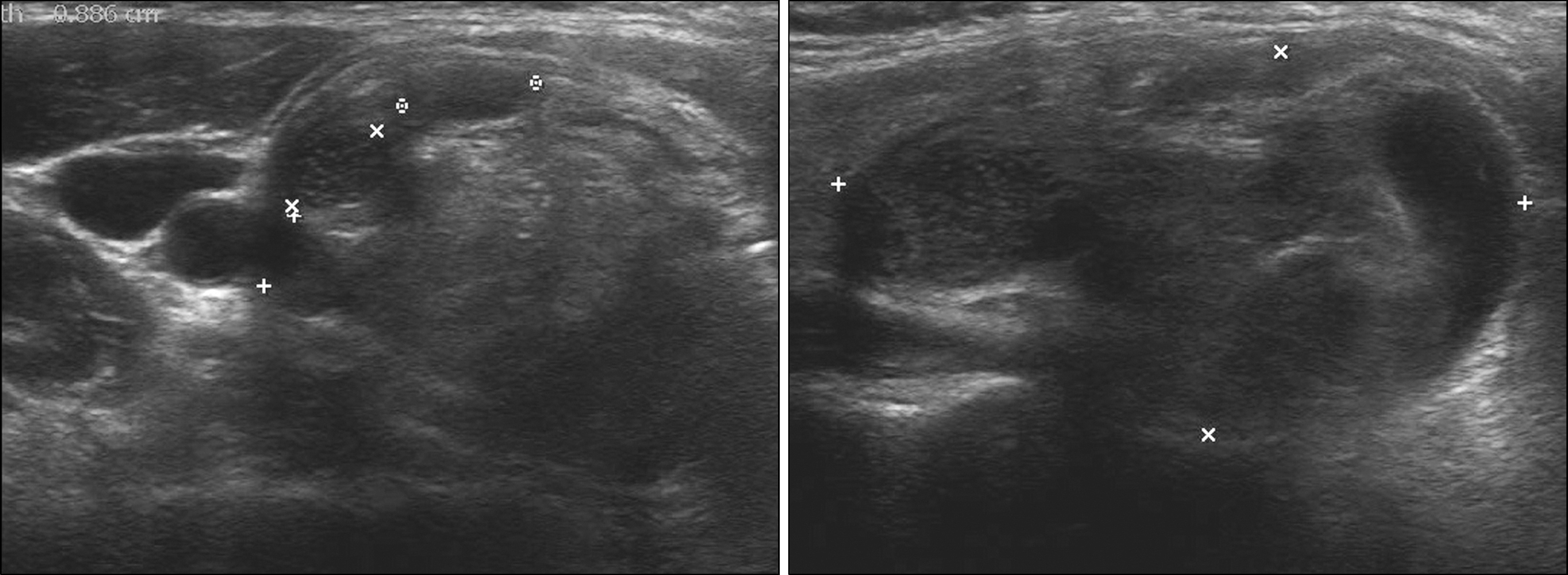
Fig. 1. Ultrasonographic scans showing right lobe was mostly occupied with a solid nodule of 44×23×25 mm with markedly inhomogeneous hypoechogenicity and hyperechoic lesion.

Fig. 2. Cut surface of teratoma demonstrated a well capsu-lated cystic mass with yellow gelatinous and grumous material admixed with many black colored hairs.

Fig. 3. Histologic findings of the benign thyroid teratoma. (A) Squamous cell epithelium and many skin appendages including hair follicles, hairs and many sebaceous glands and sweet glands are seen (H&E staining, ×50). (B) The cystic wall partly contains cartilage and bony tissue (H&E staining, ×50). (C) Mature fat tissue and smooth muscle are observed (H&E staining, ×100).(D) Nerves and vessel structures are also seen (H&E staining, ×100).
Reference
-
1). Fan SQ, Liang QC, Jiang Y. Thyroid teratoma in an 11-month-old infant. Int J Surg. 2008; 6(6):462–4.
Article
