The Clinical Characteristics of Ocular Toxocariasis in Jeju Island Using Ultra-wide-field Fundus Photography
- Affiliations
-
- 1Department of Ophthalmology, Jeju National University School of Medicine, Jeju, Korea. righthanded8282@gmail.com
- KMID: 2378626
- DOI: http://doi.org/10.3341/jkos.2017.58.5.554
Abstract
- PURPOSE
To investigate the clinical features and treatment outcomes of ocular toxocariasis in Jeju Island using ultra-wide-field fundus photography.
METHODS
We retrospectively reviewed the medical records of 40 eyes of 37 patients who were diagnosed with ocular toxocariasis based on clinical aspects and serologic tests. The quality of history-taking was assessed and peripheral blood samples were analyzed. Ocular characteristics were evaluated using ultra-wide-field fundus photography and optical coherence tomography. Changes in visual acuity and funduscopic findings after treatment were also analyzed.
RESULTS
The average age of the patients was 57.9 years and the mean Toxocara canis IgG titer was 1.979 ± 0.486. The most common fundus findings were vitreous opacity (63.6%) and granuloma (60%). Granulomas that were not initially observed within the field of view of conventional fundus photography were found using ultra-wide-field imaging in 15 eyes (62.5%). Ultra-wide-field fluorescein angiography showed peripheral vascular leakage in 16 eyes (69.6%). Treatment with oral prednisolone and albendazole resulted in average vision improvements of 0.19 ± 0.07 logMAR (p = 0.031) as well as significant improvements in anterior chamber inflammation and vitreous opacity. Combination therapy led to a significantly lower recurrence rate than prednisolone monotherapy (p = 0.049).
CONCLUSIONS
In Jeju Island, the mean Toxocara canis IgG titer of ocular toxocariasis was high. The incidences of vitreous opacity and granulomas were also high. Ultra-wide-field fundus imaging was useful for finding peripheral retinal lesions and peripheral vascular leakage that were not observed within the field of view of conventional fundus photography. Ultra-wide-field fundus imaging was valuable not only during clinical diagnosis, but also on follow-up evaluations of ocular toxocariasis. Treatment with oral prednisolone and albendazole effectively improved ocular inflammation and visual acuity and helped reduce the recurrence rate.
MeSH Terms
-
Albendazole
Anterior Chamber
Diagnosis
Fluorescein Angiography
Follow-Up Studies
Granuloma
Humans
Immunoglobulin G
Incidence
Inflammation
Medical Records
Photography*
Prednisolone
Recurrence
Retinaldehyde
Retrospective Studies
Serologic Tests
Tomography, Optical Coherence
Toxocara canis
Toxocariasis*
Uveitis
Visual Acuity
Albendazole
Immunoglobulin G
Prednisolone
Retinaldehyde
Figure
-

Figure 1 Ultra-wide-field fundus photographs and ultra-wide-field fluorescein angiographic imaging of ocular toxocariasis. (A) A granuloma with mild vitreous opacity. (B) A tractional retinal fold with localized tractional retinal detachment. (C) Diffuse peripheral vascular leakage. (D) A prominent optic disc leakage.

Figure 2 Ultra-wide-field fundus images with overlay of the Early Treatment Diabetic Retinopathy Study (ETDRS) 7-standard 30-degree fields. Small peripheral granuloma in nasal retina (A) and two peripheral granulomas in inferior retina (B) were noted in the image beyond the ETDRS 7 fields.
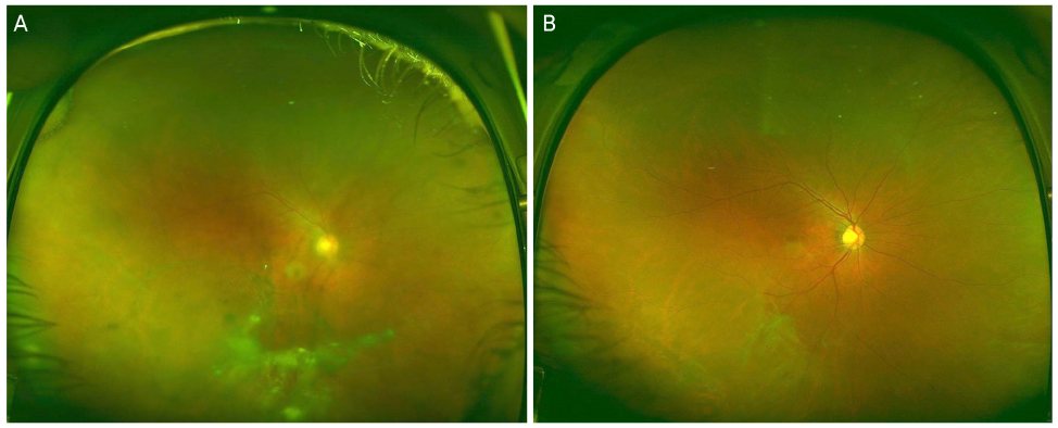
Figure 3 Ultra-wide-field fundus photographic findings of a patient with ocular toxocariasis before and after treatment of oral prednisolone with albendazole. (A) Severe vitreous opacity and multiple vitreous debris are shown. (B) The vitreous opacity was cleared up after treatment.
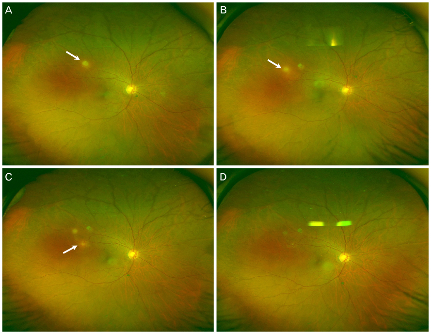
Figure 4 Ultra-wide-field fundus photographs of discontinuous migration of granuloma. (A) A round shape granuloma (white arrow) is shown. (B) After 3 months, a new granuloma (white arrow) appeared adjacent to the site of previous granuloma. (C) 1 week later, another novel granuloma (white arrow) was noted. (D) After 3 weeks, the granulomas were decreased in size.
Reference
-
1. Ahn SJ, Ryoo NK, Woo SJ. Ocular toxocariasis: clinical features, diagnosis, treatment, and prevention. Asia Pac Allergy. 2014; 4:134–141.2. Stewart JM, Cubillan LD, Cunningham ET Jr. Prevalence, clinical features, and causes of vision loss among patients with ocular toxocariasis. Retina. 2005; 25:1005–1013.3. Despommier D. Toxocariasis: clinical aspects, epidemiology, medical ecology, and molecular aspects. Clin Microbiol Rev. 2003; 16:265–272.4. Despreaux R, Fardeau C, Touhami S, et al. Ocular toxocariasis: clinical features and long-term visual outcomes in adult patients. Am J Ophthalmol. 2016; 166:162–168.5. Ahn SJ, Woo SJ, Jin Y, et al. Clinical features and course of ocular toxocariasis in adults. PLoS Negl Trop Dis. 2014; 8:e2938.6. Nicholson BP, Nigam D, Miller D, et al. Comparison of wide-field fluorescein angiography and 9-field montage angiography in uveitis. Am J Ophthalmol. 2014; 157:673–677.7. Kaines A, Tsui I, Sarraf D, Schwartz S. The use of ultra wide field fluorescein angiography in evaluation and management of uveitis. Semin Ophthalmol. 2009; 24:19–24.8. Soliman AZ, Silva PS, Aiello LP, Sun JK. Ultra-wide field retinal imaging in detection, classification, and management of diabetic retinopathy. Semin Ophthalmol. 2012; 27:221–227.9. Prasad PS, Oliver SC, Coffee RE, et al. Ultra wide-field angiographic characteristics of branch retinal and hemicentral retinal vein occlusion. Ophthalmology. 2010; 117:780–784.10. Anderson L, Friberg TR, Singh J. Ultrawide-angle retinal imaging and retinal detachment. Semin Ophthalmol. 2007; 22:43–47.11. Lee HW, Kim HC. A case of atypical acute retinal necrosis observed using ultra-wide-field imaging. J Korean Ophthalmol Soc. 2015; 56:452–457.12. Bae KW, Ahn SJ, Park KH, Woo SJ. Diagnostic value of the serum anti-toxocara IgG titer for ocular toxocariasis in patients with uveitis at a tertiary hospital in Korea. Korean J Ophthalmol. 2016; 30:258–264.13. Jin Y, Shen C, Huh S, et al. Serodiagnosis of toxocariasis by ELISA using crude antigen of toxocara canis larvae. Korean J Parasitol. 2013; 51:433–439.14. Kim YH, Huh S, Chung YB. Seroprevalence of toxocariasis amomg healthy people with eosinophilia. Korean J Parasitol. 2008; 46:29–32.15. Kwon NH, Oh MJ, Lee SP, et al. The clinical impact of toxocariasis in patients with unknown eosinophilia. Korean J Asthma Allergy Clin Immunol. 2005; 25:299–304.16. Jabs DA, Nussenblatt RB, Rosenbaum JT. Standardization of Uveitis Nomenclature (SUN) Working Group. Standardization of uveitis nomenclature for reporting clinical data Results of the First International Workshop. Am J Ophthalmol. 2005; 140:509–516.17. Nussenblatt RB, Palestine AG, Chan CC, Roberge F. Standardization of vitreal inflammatory activity in intermediate and posterior uveitis. Ophthalmology. 1985; 92:467–471.18. Sugar EA, Jabs DA, Altaweel MM, et al. Identifying a clinically meaningful threshold for change in uveitic macular edema evaluated by optical coherence tomography. Am J Ophthalmol. 2011; 152:1044–1052.e5.19. Stevenson W, Prospero Ponce CM, Agarwal DR, et al. Epiretinal membrane: optical coherence tomography-based diagnosis and classification. Clin Ophthalmol. 2016; 10:527–534.20. Kang HM, Lee SC. Long-term progression of retinal vasculitis in Behçet patients using a fluorescein angiography scoring system. Graefes Arch Clin Exp Ophthalmol. 2014; 252:1001–1008.21. Kim M, Kwon HJ, Choi EY, et al. Correlation between fluorescein angiographic findings and visual acuity in Behçet retinal vasculitis. Yonsei Med J. 2015; 56:1087–1096.
- Full Text Links
-
- Actions
-
Cited
- CITED
-
- Close
- Share
-
- Similar articles
-
- The Extent of Silicone Oil Emulsification Revealed by Ultra-wide-field Fundus Photography and Optical Coherence Tomography
- Diagnostic Availability of Ultra-Wide-field Fundus Imaging in Korean Patient with Retinal Break
- Ultra-wide Field Fundus Photography Using Eye Steering Technique in Patients with Symptomatic Posterior Vitreous Detachment
- Serial Follow-up of Commotio Retinae Using Ultra-wide Field Imaging
- Utility of Ultra-wide Fundus Photography in Patients with Acute Blunt Ocular Trauma
