Localization of mandibular canal and assessment of the remaining alveolar bone in posterior segment of the mandible with single missing tooth using cone-beam computed tomography: a cross sectional comparative study
- Affiliations
-
- 1Division of Periodontics, Ministry of Health, College of Dentistry, Qassim Private Colleges, Buraydah, Kingdom of Saudi Arabia.
- 2Division of Radiology, Department of Oral, Basic and Clinical Sciences, College of Dentistry, Qassim Private Colleges, Buraydah, Kingdom of Saudi Arabia. saif.fahad111@gmail.com
- KMID: 2377013
- DOI: http://doi.org/10.5125/jkaoms.2017.43.2.100
Abstract
OBJECTIVES
Localization of the mandibular canal (MC) and measurement of the height and width of the available alveolar bone at the proposed implant site in the posterior segment of the mandible using cone-beam computed tomography (CBCT) in patients with a single missing tooth.
MATERIALS AND METHODS
A cross-sectional study was performed where CBCT scans of the patients with a single missing tooth in the posterior segment of the mandible"”premolar, I (1st) molar, and II (2nd) molar were used. The scans were assessed using OnDemand3D software (version 1.0; CyberMed Inc., Seoul, Korea) for localization of the MC asnd remaining alveolar bone both vertically (from the superior position of the MC to the crest of the alveolar ridge) and horizontally (buccolingual, 3 mm below the crest of the alveolar ridge). The findings were statistically analyzed using independent t-test.
RESULTS
A total of 120 mandibular sites (40 sites for each of the three missing premolar, I molar, and II molar) from 91 CBCT scans were analyzed. The average heights (from the alveolar crest to the superior margin of the MC) at the premolar, I molar, and II molar areas were 15.19±2.12 mm, 14.53±2.34 mm, and 14.21±2.23 mm, respectively. The average widths, measured 3 mm below the crest of the alveolar ridge, at the premolar, I molar, and II molar areas were 6.22±1.96 mm, 6.51±1.75 mm, and 7.60±2.08 mm, respectively. There was no statistically significant difference between males and females regarding the vertical and horizontal measurements of the alveolar ridges.
CONCLUSION
In the study, the measurements were averaged separately for each of the single missing teeth (premolar, I molar, or II molar), giving more accurate information for dental implant placement.
MeSH Terms
Figure
-

Fig. 1 Axial cut of the lower left arch showing a curved line drawn through the adjacent teeth roots to produce a panoramic view.
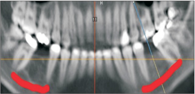
Fig. 2 Panoramic view showing the tracing of the mandibular canal at the proposed implant placement site.
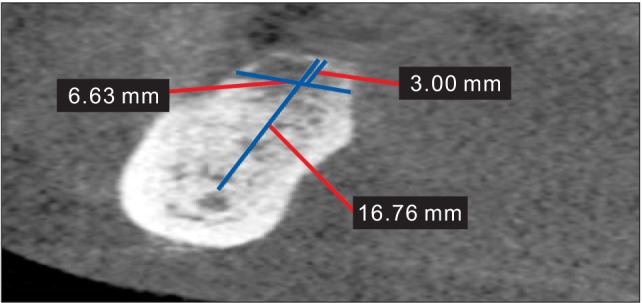
Fig. 3 Cross-sectional view showing the remaining alveolar bone of the edentulous space for tooth #46, assessed in regard to the vertical height from the superior position of the mandibular canal to the crest of the alveolar ridge (16.76 mm). Bucco-lingual width was assessed 3.00 mm below the crest of the alveolar ridge (6.63 mm).
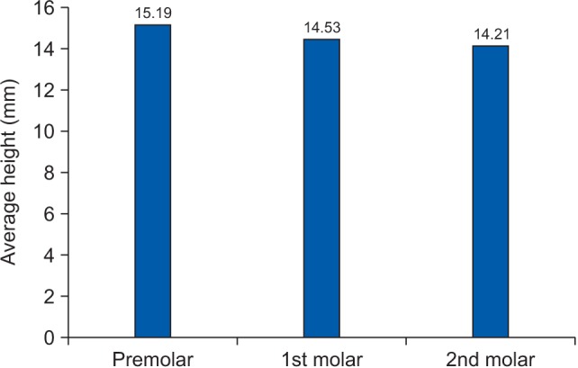
Fig. 4 Average height in mm from the alveolar crest to the superior position of the mandibular canal at the premolar, I (1st) molar, and II (2nd) molar areas.

Fig. 5 Average horizontal width in mm of the alveolar ridge in the mandible at the premolar, I (1st) molar, and II (2nd) molar areas.
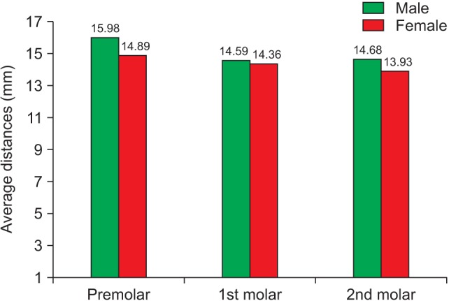
Fig. 6 Average distances in mm from the alveolar crest to the superior position of the mandibular canal based on gender.
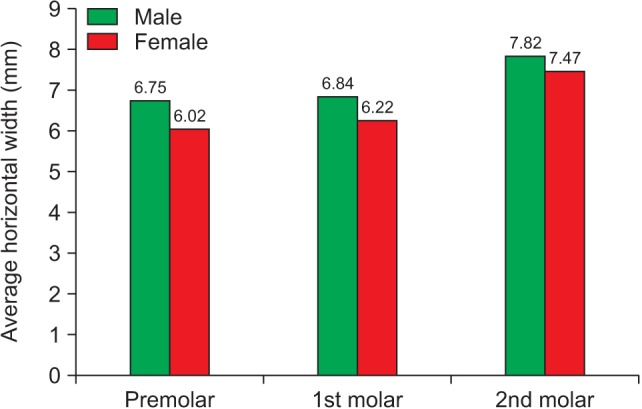
Fig. 7 Average horizontal width in mm of the alveolar ridge in the mandible based on gender.
Reference
-
1. Khalifa N, Allen PF, Abu-bakr NH, Abdel-Rahman ME. Factors associated with tooth loss and prosthodontic status among Sudanese adults. J Oral Sci. 2012; 54:303–312. PMID: 23221155.
Article2. Cahen PM, Frank RM, Turlot JC. A survey of the reasons for dental extractions in France. J Dent Res. 1985; 64:1087–1093. PMID: 2862168.
Article3. Chauncey HH, Muench ME, Kapur KK, Wayler AH. The effect of the loss of teeth on diet and nutrition. Int Dent J. 1984; 34:98–104. PMID: 6588038.4. Sullivan RM. Implant dentistry and the concept of osseointegration: a historical perspective. J Calif Dent Assoc. 2001; 29:737–745. PMID: 11806052.5. Araújo MG, Lindhe J. Dimensional ridge alterations following tooth extraction. An experimental study in the dog. J Clin Periodontol. 2005; 32:212–218. PMID: 15691354.
Article6. Eufinger H, König S, Eufinger A. The role of alveolar ridge width in dental implantology. Clin Oral Investig. 1997; 1:169–177.
Article7. Eufinger H, König S, Eufinger A, Machtens E. Significance of the height and width of the alveolar ridge in implantology in the edentulous maxilla. Analysis of 95 cadaver jaws and 24 consecutive patients. Mund Kiefer Gesichtschir. 1999; 3(Suppl 1):S14–S18. PMID: 10414076.8. Kim SG. Clinical complications of dental implants. In : Turkyilmaz I, editor. Implant dentistry: a rapidly evolving practice. Rijeka: InTech;2011. DOI: 10.5772/17262.9. Moslehifard E, Nikzad S, Geraminpanah F, Mahboub F. Full-mouth rehabilitation of a patient with severely worn dentition and uneven occlusal plane: a clinical report. J Prosthodont. 2012; 21:56–64. PMID: 21981748.
Article10. McCrea SJ. Pre-operative radiographs for dental implants: are selection criteria being followed? Br Dent J. 2008; 204:675–682. PMID: 18587363.11. Reddy MS, Mayfield-Donahoo T, Vanderven FJ, Jeffcoat MK. A comparison of the diagnostic advantages of panoramic radiography and computed tomography scanning for placement of root form dental implants. Clin Oral Implants Res. 1994; 5:229–238. PMID: 7640337.
Article12. Hanazawa T, Sano T, Seki K, Okano T. Radiologic measurements of the mandible: a comparison between CT-reformatted and conventional tomographic images. Clin Oral Implants Res. 2004; 15:226–232. PMID: 15008935.13. Scarfe WC, Farman AG, Sukovic P. Clinical applications of cone-beam computed tomography in dental practice. J Can Dent Assoc. 2006; 72:75–80. PMID: 16480609.14. Benavides E, Rios HF, Ganz SD, An CH, Resnik R, Reardon GT, et al. Use of cone beam computed tomography in implant dentistry: the International Congress of Oral Implantologists consensus report. Implant Dent. 2012; 21:78–86. PMID: 22382748.15. Angelopoulos C, Thomas SL, Hechler S, Parissis N, Hlavacek M. Comparison between digital panoramic radiography and cone-beam computed tomography for the identification of the mandibular canal as part of presurgical dental implant assessment. J Oral Maxillofac Surg. 2008; 66:2130–2135. PMID: 18848113.
Article16. Kim ST, Hu KS, Song WC, Kang MK, Park HD, Kim HJ. Location of the mandibular canal and the topography of its neurovascular structures. J Craniofac Surg. 2009; 20:936–939. PMID: 19461335.
Article17. Juodzbalys G, Wang HL, Sabalys G. Anatomy of mandibular vital structures. Part I: mandibular canal and inferior alveolar neurovascular bundle in relation with dental implantology. J Oral Maxillofac Res. 2010; 1:e2.
Article18. Shahin KA, Chatra L, Shenai P. Stature estimating the location of maxillary sinus and mandibular canal. N Am J Med Sci. 2012; 4:586–589. PMID: 23181232.
Article19. Waltrick KB, Nunes de, Corrêa M, Zastrow MD, Dutra VD. Accuracy of linear measurements and visibility of the mandibular canal of cone-beam computed tomography images with different voxel sizes: an in vitro study. J Periodontol. 2013; 84:68–77. PMID: 22390549.
Article20. Kilic C, Kamburoğlu K, Ozen T, Balcioglu HA, Kurt B, Kutoglu T, et al. The position of the mandibular canal and histologic feature of the inferior alveolar nerve. Clin Anat. 2010; 23:34–42. PMID: 19918867.
Article21. Pramstraller M, Farina R, Franceschetti G, Pramstraller C, Trombelli L. Ridge dimensions of the edentulous posterior maxilla: a retrospective analysis of a cohort of 127 patients using computerized tomography data. Clin Oral Implants Res. 2011; 22:54–61. PMID: 20831759.
Article22. de Oliveira Júnior MR, Saud AL, Fonseca DR, De-Ary-Pires B, Pires-Neto MA, de Ary-Pires R. Morphometrical analysis of the human mandibular canal: a CT investigation. Surg Radiol Anat. 2011; 33:345–352. PMID: 20677005.
Article23. Levine MH, Goddard AL, Dodson TB. Inferior alveolar nerve canal position: a clinical and radiographic study. J Oral Maxillofac Surg. 2007; 65:470–474. PMID: 17307595.
Article24. Watanabe H, Mohammad Abdul M, Kurabayashi T, Aoki H. Mandible size and morphology determined with CT on a premise of dental implant operation. Surg Radiol Anat. 2010; 32:343–349. PMID: 19812884.
Article25. Frei C, Buser D, Dula K. Study on the necessity for cross-section imaging of the posterior mandible for treatment planning of standard cases in implant dentistry. Clin Oral Implants Res. 2004; 15:490–497. PMID: 15248885.
Article26. Braut V, Bornstein MM, Lauber R, Buser D. Bone dimensions in the posterior mandible: a retrospective radiographic study using cone beam computed tomography. Part 1--analysis of dentate sites. Int J Periodontics Restorative Dent. 2012; 32:175–184. PMID: 22292147.
- Full Text Links
-
- Actions
-
Cited
- CITED
-
- Close
- Share
-
- Similar articles
-
- Assessment of the relationship between the mandibular third molar and the mandibular canal using panoramic radiograph and cone beam computed tomography
- Comparative evaluation of computed tomography for dental implants on the mandibular edentulous area
- Sex determination by radiographic localization of the inferior alveolar canal using cone-beam computed tomography in an Egyptian population
- Radiographic evaluation of the course and visibility of the mandibular canal
- Cone beam CT findings of retromolar canals: Report of cases and literature review
