Prog Med Phys.
2016 Dec;27(4):241-249. 10.14316/pmp.2016.27.4.241.
Evaluations of the Space Dose and Dose Reductions in Patients and Practitioners by Using the C-arm X-ray Tube Shielding Devices Developed in Our Laboratory
- Affiliations
-
- 1Department of Medical Physics, Kyonggi University, Suwon, Korea.
- 2Korea Institute of Radiological and Medical Sciences, Seoul, Korea. haijo@kirams.re.kr
- 3Department of Radiological Science, Sin-Gu University, Seongnam, Korea.
- KMID: 2376559
- DOI: http://doi.org/10.14316/pmp.2016.27.4.241
Abstract
- The present study used a digital angiography x-ray device to measure the space dose and exposure dose of patients and practitioners using x-ray tube shielding devices developed in our laboratory. The intent of the study was to reduce the space dose within the test room, and to reduce the exposure dose of patients and practitioners. The patient and practitioner exposure doses were measured in five configurations in a human body model. The glass dosimeter was placed on the eye lenses, thyroid glands, left shoulder, right shoulder, and gonads. The beam was collimated at full size and at a 48% reduction for a comparative analysis of the measurements. The space dose was measured with an ion chamber at distances of 50 cm, 100 cm, and 150 cm from the x-ray tube under the following conditions: no shielding device; a shielding device made of 3-mm-thick lead (Pb) [Pb 3 mm shield], and a shielding device made of 3-mm-thick Pb (outside) and 3-mm-thick aluminum (Al) (inside) [Pb 3 mm+Al 3 mm shield]. The absorbed dose was the lowest when the 3-mm-thick Pb+3-mm-thick Al shield was used. For measurements made with collimated beams with a 48% reduction, the dose was the lowest at 154 µGy when the 3-mm-thick Pb+3-mm-thick Al shield was used, and was 9 µGy lower than the measurements made with no shielding device. If the space dose can be reduced by 20% in all situations where the C-arm is employed by using the x-ray tube shielding devices developed in our laboratory, this is expected to play an important role in reducing the annual exposure dose for patients, practitioners, and assistants.
Keyword
MeSH Terms
Figure
-
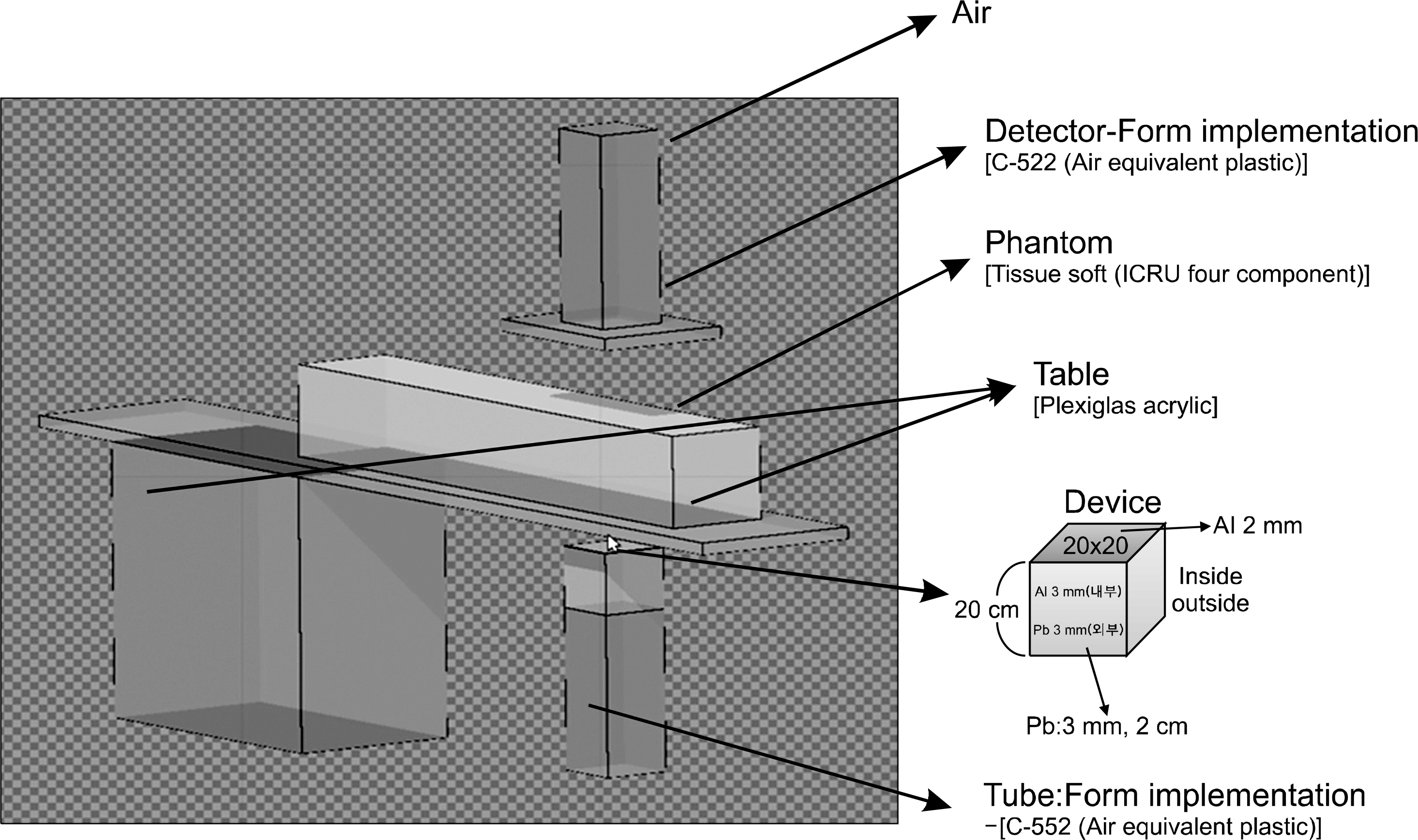
Fig. 1. Geometry design for Monte Carlo simulation.
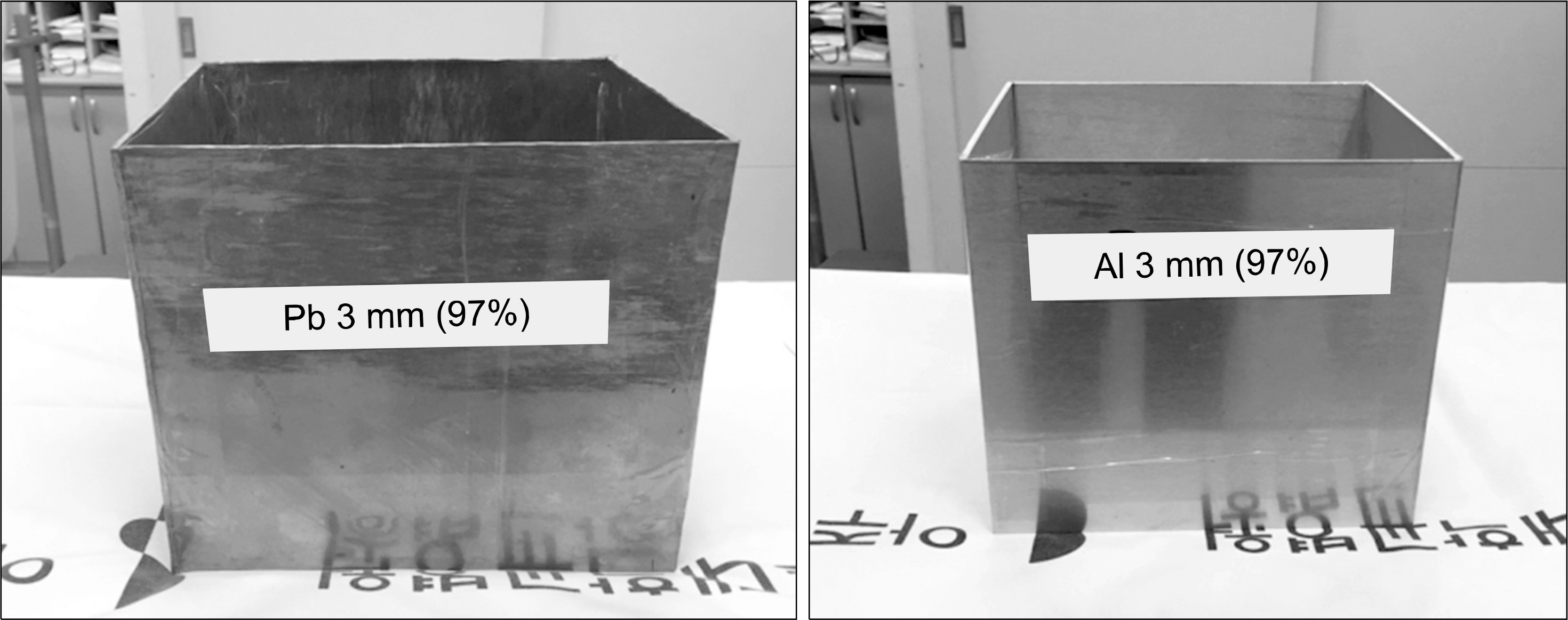
Fig. 2. 3-mm-thick Pb (x-ray tube shield) and 3-mm-thick Al (x-ray tube double shield).
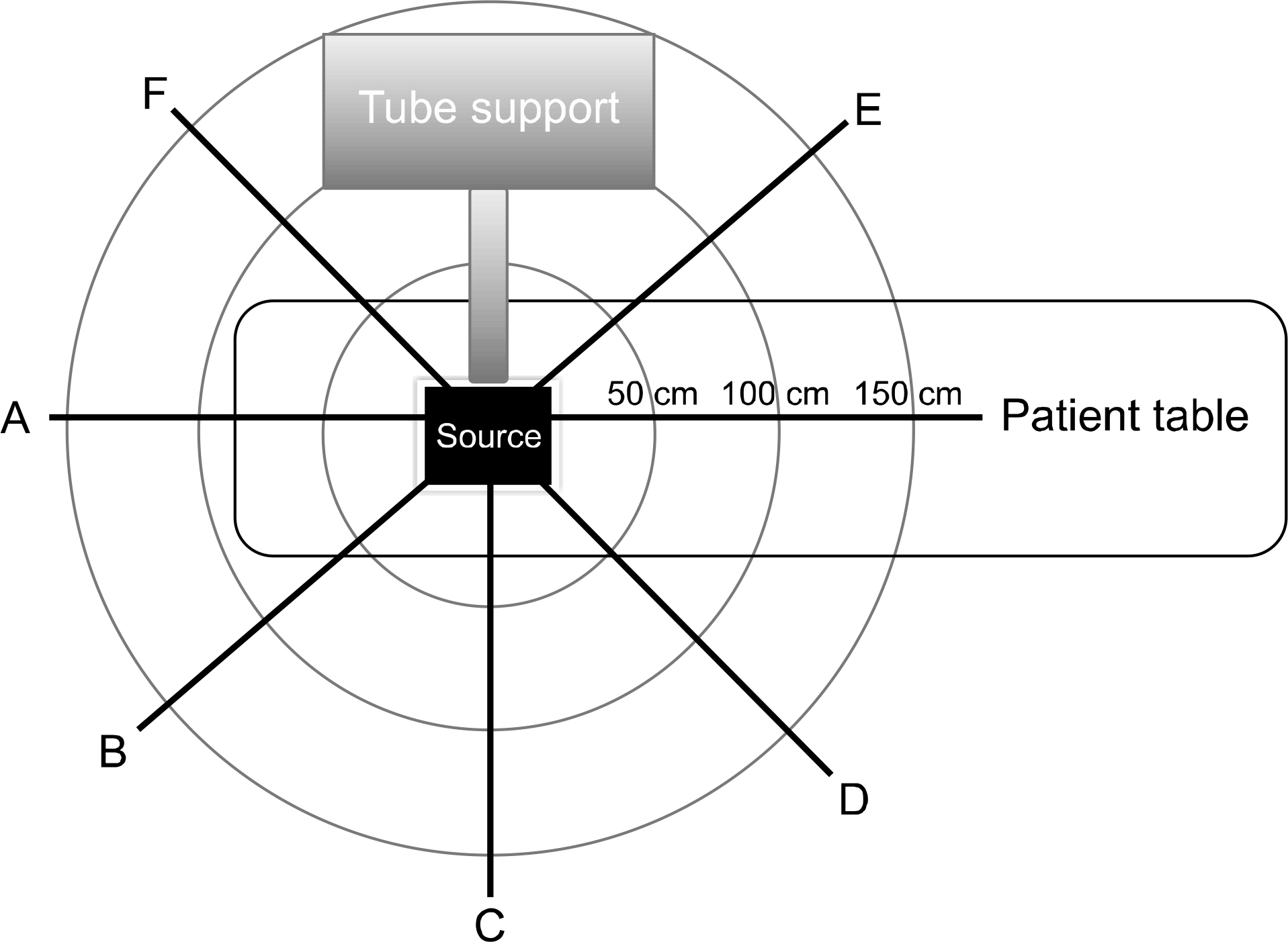
Fig. 3. Schematic diagram of the measurement of six directions (A-F).

Fig. 4. Arbitrary dose unit based on the distance in the Z axis.
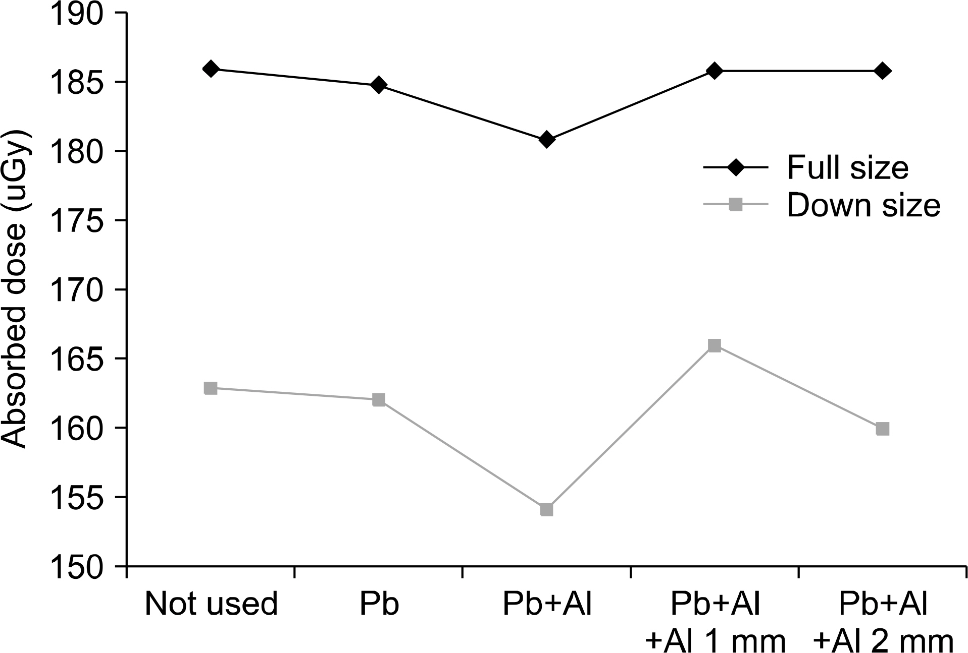
Fig. 5. Comparison of the absorbed dose of the full size & down-sized beam collimations in the phantom.
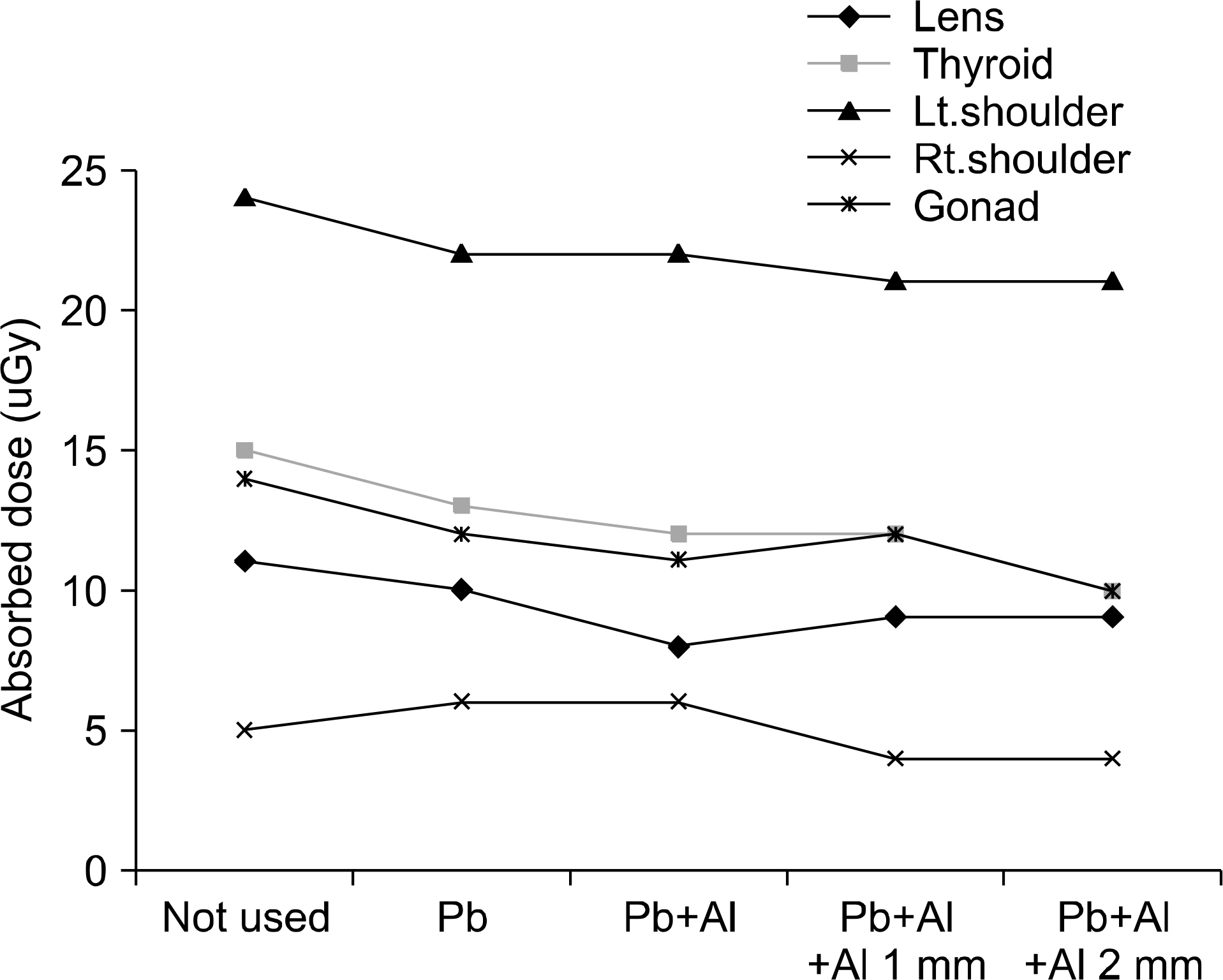
Fig. 6. Comparison of the absorbed doses of the lenses, thyroid, left, right shoulder, and gonads depending on the presence or absence of shielding.
Reference
-
1. NCRP report 160. Ionizing radiation exposure of the population of the United States, medical exposure: are we doing less with more, and is there a role for health physicists. Health Phys, Schauer DA, Linton OW. 2009.2. Woo Kyoung Jeong. Radiation exposure and its reduction in the fluoroscopic examination and fluoroscopy Guided interventional radiology, Korea Medical Association. 54(12):1269–1270.3. Schueler BA. Operator shielding: how and why. Tech Vasc Interv Radiol. 13:167–171. 2010.
Article4. NCRP report 116. Limitation of exposure to ionizing radiation. National Council on Radiation Protection and Measurements, Bethesda. 1993.5. http://sizekorea.kats.go.kr6. Vano E, Gonzalez L, Beneytez F, et al. Lens injuries induced by occupational exposure in non-optimised interventional radiology laboratories. Brit J Radiol Vol. 71:728–733. 1998.7. Vano E, Gonzalez L, Guibelalde E, et al. Radiation exposure to medical staff in interventional and cardiac radiology. Brit J Radiol Vol. 71:954–960. 1998.8. Report. Occupational Radiation Exposure in Diagnostic Radiology in Korea. Korea Food & Drug Administration, Medical Institutions, Seung Hee Kim. 2008.9. Brateman L. Radiation safety considerations for diagnostic radiology personnel. Radiographics. 19:1037–1055. 1999.
- Full Text Links
-
- Actions
-
Cited
- CITED
-
- Close
- Share
-
- Similar articles
-
- A Study on Dose Distribution around Fletcher-Suit Colpostat Containing Cs-137 Source by a Computer
- The Efficiency of Radiation Shielding Sheet to Reduce Radiation Exposure during C-arm Fluoroscopy
- Dose distribution of high energy electron by shielding material
- Dose Distribution for Eye Shielding Block in 6 MV Photon Beam Therapy
- Dose Distribution in the Brain in Radiotherapy of Whole Barin
