Gastric Adenocarcinoma with Prostatic Metastasis
- Affiliations
-
- 1Department of Radiation Oncology, Regional Cancer Centre, Thiruvananthapuram, India.
- 2Department of Medical Oncology, Regional Cancer Centre, Thiruvananthapuram, India. dranooptm@yahoo.co.in
- 3Department of Pathology, Regional Cancer Centre, Thiruvananthapuram, India.
- 4Department of Surgical Oncology, Regional Cancer Centre, Thiruvananthapuram, India.
Abstract
- Metastasis of gastric adenocarcinoma to the prostate gland is extremely rare. Herein, we report a case of gastric adenocarcinoma in a 56-year-old man with prostatic metastasis diagnosed through the analysis of biopsy specimens from representative lesions in the stomach and prostate gland. Immunohistochemistry of the prostatic tissue showed positive staining for cytokeratin 7 and negative staining for prostate-specific antigen (PSA), whereas the serum PSA level was normal, confirming the diagnosis of prostatic metastasis from carcinoma of the stomach.
MeSH Terms
Figure
-
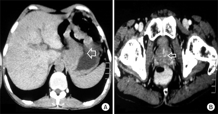
Fig. 1 (A) Computed tomography of the abdomen and pelvis showing mural wall thickening involving the gastroesophageal junction and extending to the lesser curvature of the stomach. (B) Computed tomography of the abdomen and pelvis with arrow showing an enlarged prostatic mass.
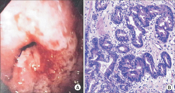
Fig. 2 (A) Upper gastrointestinal endoscopy showing an ulceroinfiltrative growth at the gastroesophageal junction along the lesser curvature of the stomach. (B) Histopathology slides (H&E, ×200) showing moderate-to-poorly differentiated gastric adenocarcinoma with gastric mucosa and ulceration, and cells arranged in a glandular pattern, signet ring cells, and pools of mucin.
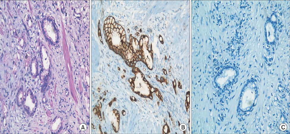
Fig. 3 (A) Tru-cut biopsy slides (H&E, ×200) of the prostate gland showing infiltration by small and large glands, lined by tall columnar/cuboidal mucinous cells. (B) Immunohistochemical analysis (×400) of prostatic tissue showing cytokeratin 7 positivity. (C) Immunohistochemical analysis (×400) of prostatic tissue showing prostate-specific antigen negativity.
Reference
-
1. Zhang P, Zheng Y, Ran H, Leng Z, Wang Z. Case report: gastric adenocarcinoma metastatic to the prostate gland. J Radiol Case Rep. 2010; 4:35–38.
Article2. Borum ML, Chen HC. Gastric adenocarcinoma metastatic to the prostate gland: a rare case and review of the literature. Dig Dis Sci. 2001; 46:2658–2659.3. Bates AW, Baithun SI. Secondary solid neoplasms of the prostate: a clinico-pathological series of 51 cases. Virchows Arch. 2002; 440:392–396.
Article4. Epstein JI. PSA and PAP as immunohistochemical markers in prostate cancer. Urol Clin North Am. 1993; 20:757–770.
Article
- Full Text Links
-
- Actions
-
Cited
- CITED
-
- Close
- Share
-
- Similar articles
-
- Cutaneous Metastases from Prostatic Adenocarcinoma
- A Case of Brain Metastasis from Prostatic Adenocarcinoma Which Showed Remarkable Effect in Combined Chemotherapy
- Fine Needle Aspiration Cytology of Metastatic Prostatic Adenocarcinoma, Pseudohyperplastic Variant
- Prostatic Adenocarcinoma Presenting as an Abdominal Mass
- A Case of Epididymal Metastasis from Mucinous Adenocarcinoma of the prostate
