J Gastric Cancer.
2012 Dec;12(4):258-261.
Ewing's Sarcoma of the Stomach; Rare Case of Ewing's Sarcoma and Suggestion of New Treatment Strategy
- Affiliations
-
- 1Department of Surgery, Chosun University Hospital, School of Medicine, Chosun University, Gwangju, Korea. sungsoo73@chosun.ac.kr
- 2Department of Pathology, Chosun University Hospital, School of Medicine, Chosun University, Gwangju, Korea.
Abstract
- Ewing's sarcoma is a neoplasm of the undifferenciated small round cells, which generally affects the bone and deep soft tissues of children and adolescents. We present a case of gastric Ewing's sarcoma; a 35-year-old female who had no symptoms. While she was at a routine medical checkup, a protruding mass in her gastric antrum was incidentally found on esophagogastroduodenoscopy. Endoscopic ultrasonogram showed a submucosal mass on the same lesion and a laparosopic wedge resection was done. Pathologic gross findings showed a granular grape appearance tissue and histoloigc examination revealed a small round cell tumor with CD 99 immunoexpression positive. In general, a combined modality therapy for Ewing's sarcoma such as surgical resection with chemotherapy, is accepted as an effective method. However, this patient had no adjuvant chemotherapy after surgery and she has no recurrence for eleven months.
MeSH Terms
Figure
-
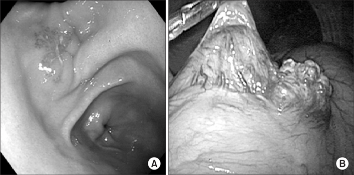
Fig. 1 (A) Endoscopic view. A 2.5 cm sized protruding mass with central depression and erythematous change is noted in the anterior wall of proximal antrum just below angle. (B) Intraoperative laparoscopic view. Granular grape appearance mass is noted on laparoscopic view.
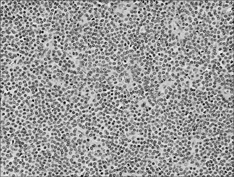
Fig. 2 Relativley monomorphic small cells with round or oval nuclei with well defined nuclear membrane (H&E, ×200).
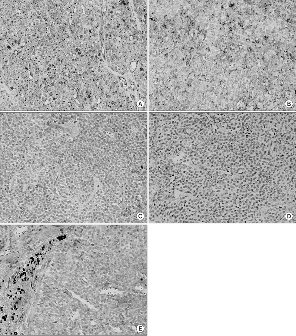
Fig. 3 (A) Immunohistology. CD99 (+) (×400). (B) Immunohistology. Synaptophysin (+) (×400). (C) Immunohistology. Chromogranin A (-) (×400). (D) Immunohistology. Cytokeratin (-) (×400). (E) Immunohistology. Desmin (-) (×400).
Reference
-
1. El Weshi A, Allam A, Ajarim D, Al Dayel F, Pant R, Bazarbashi S, et al. Extraskeletal Ewing's sarcoma family of tumours in adults: analysis of 57 patients from a single institution. Clin Oncol (R Coll Radiol). 2010. 22:374–381.
Article2. Iwamoto Y. Diagnosis and treatment of Ewing's sarcoma. Jpn J Clin Oncol. 2007. 37:79–89.
Article3. Soulard R, Claude V, Camparo P, Dufau JP, Saint-Blancard P, Gros P. Primitive neuroectodermal tumor of the stomach. Arch Pathol Lab Med. 2005. 129:107–110.
Article4. Czekalla R, Fuchs M, Stölzle A, Nerlich A, Poremba C, Schaefer KL, et al. Peripheral primitive neuroectodermal tumor of the stomach in a 14-year-old boy: a case report. Eur J Gastroenterol Hepatol. 2004. 16:1391–1400.
Article5. Colovic RB, Grubor NM, Micev MT, Matic SV, Atkinson HD, Latincic SM. Perigastric extraskeletal Ewing's sarcoma: a case report. World J Gastroenterol. 2009. 15:245–247.
Article6. Shek TW, Chan GC, Khong PL, Chung LP, Cheung AN. Ewing sarcoma of the small intestine. J Pediatr Hematol Oncol. 2001. 23:530–532.
Article7. Adair A, Harris SA, Coppen MJ, Hurley PR. Extraskeletal Ewings sarcoma of the small bowel: case report and literature review. J R Coll Surg Edinb. 2001. 46:372–374.8. Kie JH, Lee MK, Kim CJ, Lee K, Kwon KW, Yang WI. Primary Ewing's sarcoma of the suodenum: a case report. Int J Surg Pathol. 2003. 11:331–337.
Article9. Lewis TB, Coffin CM, Bernard PS. Differentiating Ewing's sarcoma from other round blue cell tumors using a RT-PCR translocation panel on formalin-fixed paraffin-embedded tissues. Mod Pathol. 2007. 20:397–404.
Article10. Tanas MR, Rubin BP, Tubbs RR, Billings SD, Downs-Kelly E, Goldblum JR. Utilization of fluorescence in situ hybridization in the diagnosis of 230 mesenchymal neoplasms: an institutional experience. Arch Pathol Lab Med. 2010. 134:1797–1803.
Article
