Myelomatous Ascites as an Initial Manifestation of Extramedullary Involvement of Multiple Myeloma
- Affiliations
-
- 1Department of Radiology, Soonchunhyang University Bucheon Hospital, Bucheon, Korea. radhklee@naver.com
- 2Department of Pathology, Soonchunhyang University Bucheon Hospital, Bucheon, Korea.
- 3Department of Internal Medicine, Soonchunhyang University Bucheon Hospital, Bucheon, Korea.
- KMID: 2371684
- DOI: http://doi.org/10.3348/jksr.2017.76.3.187
Abstract
- Multiple myeloma is a common hematological malignancy. Aggressive myeloma invades the organs outside the bone marrow, lymph, or reticuloendothelial systems. Among the extramedullary involvements of multiple myeloma, myelomatous ascites are extremely rare and are associated with a poor prognosis. We describe a case of myelomatous ascites as an initial manifestation of extramedullary involvement of multiple myeloma in 39-year-old patient. The patient was treated with high-dose chemotherapy, but extensive extramedullary involvement progressed, and the patient expired approximately five months after the initial detection of ascites.
MeSH Terms
Figure
-
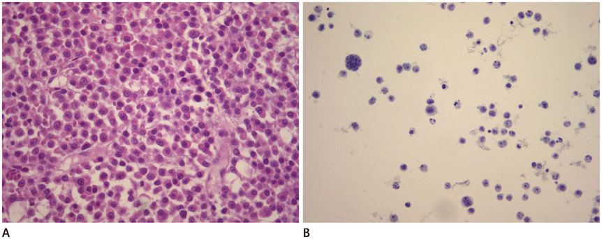
Fig. 1 Histopathology of bone marrow biopsy and ascitic fluid cytology. A. Bone marrow biopsy shows diffuse infiltration by plasma cells with moderate nuclear pleomorphism, abundant amphophilic cytoplasm, and occasional prominent nucleoli (H&E, × 400). B. Ascitic fluid cytology shows numerous large and atypical plasma cells (Pap, × 200). H&E = hematoxylin and eosin

Fig. 2 A 39-year-old male with multiple myeloma. A. In initial abdominal CT scan, take 7 days after visiting the hospital, showing a small number of ascites in the pelvic cavity with suspicious peritoneal thickening (arrows) B-D. A CT scan obtained after 3 months showing a markedly increased number of ascites with mass-like enhanced thickening of the peritoneum (arrows in B), infiltrative soft tissue attenuation encasing both kidneys and ureters (arrows in C), and homogeneously enhanced wall thickening of small bowel loops (arrows in D).
Reference
-
1. Sanal SM, Yaylaci M, Mangold KA, Pantazis CG. Extensive extramedullary disease in myeloma. An uncommon variant with features of poor prognosis and dedifferentiation. Cancer. 1996; 77:1298–1302.2. Alacacioglu L, Turgut N, Piskin O, Ali Ozcan M, Hayri Ozsan G, Demirkan F, et al. Multiple myeloma presenting as massive ascites. J BUON. 2009; 14:307–308.3. Attwell A, Dee E, Russ P, Nash R, Shah R. Multiple myeloma involving the porta hepatis and peritoneum causing biliary obstruction and malignant ascites. Dig Dis Sci. 2005; 50:1068–1071.4. Shimizu M, Tsurumi H, Hara T, Fukutomi Y, Moriwaki H. [Multiple myeloma presenting as massive ascites]. Rinsho Ketsueki. 1999; 40:515–517.5. Greer JP, Pinson RD, Russell WG, Keith TA, Collins RD, Graber SE. Malignant plasmacytic ascites. A report of two cases and a review of the literature. Cancer. 1985; 56:2001–2004.6. Karp SJ, Shareef D. Ascites as a presenting feature multiple myeloma. J R Soc Med. 1987; 80:182–184.7. Ohmoto A, Kohno M, Yasukawa K, Matsuyama R. [Massive ascites as an initial sign, plasmacytoma of the cervical supine, and hyperammonemic consciousness disturbance in a patient with biclonal type multiple myeloma]. Rinsho Ketsueki. 1996; 37:346–351.8. Alegre A, Martínez-Chamorro C, Fernández-Rañada JM. Massive myelomatous ascites responsive to VAD chemotherapy and autologous stem cell transplantation. Bone Marrow Transplant. 1999; 24:343–344.9. Jackson SR, Hawkins TE, Green GJ, Carter JM, Romeril KR. Plasmacytic ascites responsive to multiagent chemotherapy. Am J Hematol. 1992; 41:298.10. Lake G, Schade RR, Van Thiel DH. Extrahepatic biliary tract obstruction due to plasmacytoma. J Clin Gastroenterol. 1983; 5:273–276.
- Full Text Links
-
- Actions
-
Cited
- CITED
-
- Close
- Share
-
- Similar articles
-
- A case of multiple myeloma with ascites
- A Case of Multiple Myeloma with Multiple Intrahepatic Extramedullary Plasmacytomas
- A Case of Maxillary Sinus Involvement in Multiple Myeloma
- Nonsecretory Multiple Myeloma associated with Immune Thrombocytopenia and Complicated by Malignant Ascites
- A Case of Cutaneous Plasmacytoma Treated with Thalidomide
