A Rare Case of Angioleiomyoma Arising in the Subglottic Area to Upper Trachea of a Patient with Underlying Asthma
- Affiliations
-
- 1Department of Hospital Pathology, Seoul St. Mary's Hospital, College of Medicine, The Catholic University of Korea, Seoul, Korea. leekyoyo@catholic.ac.kr
- 2Division of Pulmonary, Allergy and Critical Care Medicine, Department of Internal Medicine, Seoul St. Mary's Hospital, College of Medicine, The Catholic University of Korea, Seoul, Korea.
- KMID: 2367685
- DOI: http://doi.org/10.4132/jptm.2016.06.21
Abstract
- Angioleiomyoma is a rare disease that is histologically characterized by smooth muscle cells arranged around vascular spaces. Although angioleiomyomas occur rarely in the head and neck region, they can cause various symptoms according the site involved. Here, we present a 44-year-old male patient with a 15-year history of asthma, who presented with recent onset of chest discomfort, globus sensation and throat pain. Medication was not effective in relieving his symptoms, and further evaluation revealed a polypoid ovoid mass, almost obstructing the airway at the border of the larynx and upper trachea on chest computed tomography. The mass was completely resected via a rigid bronchoscopy procedure. Histopathologic examination revealed that the excised mass was angioleiomyoma, which was immunohistochemically positive for smooth muscle actin and negative for desmin.
Keyword
MeSH Terms
Figure
-
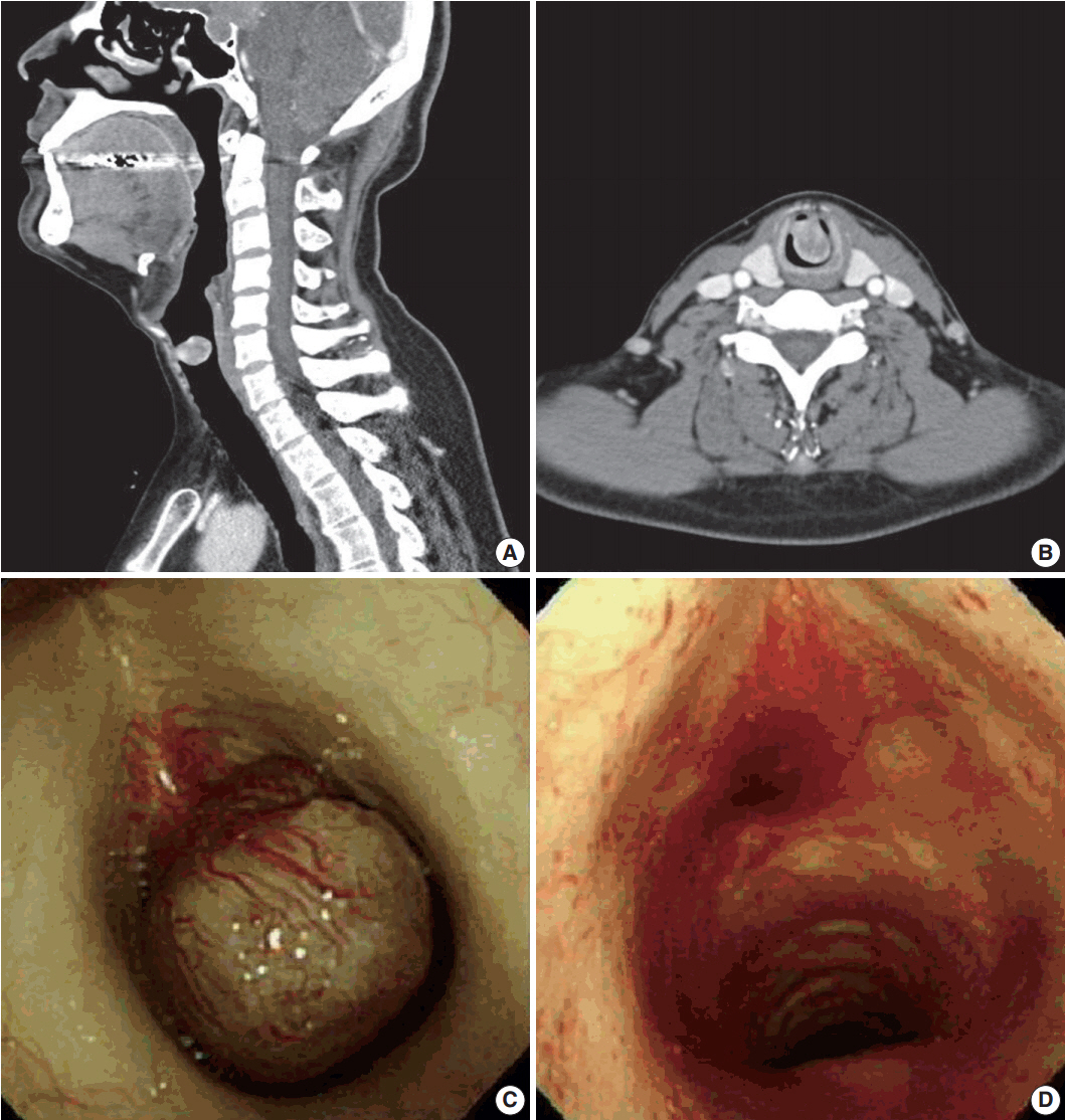
Fig. 1. Neck computed tomography (CT) and bronchoscopic findings. (A, B) Neck CT reveals a 2.0×1.5-cm-sized, round exophytic lesion at the border of the subglottic area and upper trachea. (C) Bronchoscopic finding during the operation, showing an ovoid and polypoid mass located 1 cm below the vocal cord, attached to the anterior wall of the trachea by a stalk-like structure. (D) Bronchoscopic view of the same lesion after the excision; the mass is resected by snare without complications.
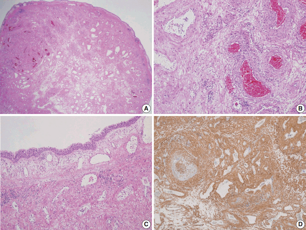
Fig. 2. Histopathologic findings of the mass. (A) Low-power view shows an ovoid and polypoid mass, consisting of well differentiated smooth muscle cells with intervening vascular channels and covered by respiratory epithelium. (B) Variable venous lumens, surrounded by muscular coats with dense to relatively loose intervascular smooth muscle cells are noted. (C) The surface is covered with ciliated pseudostratified columnar bronchial epithelium. Thin walled vessels are abundantly placed in the lamina propria, some of which have an ill-defined muscular coat. (D) Immunohistochemically, the tumor cells are positive for smooth muscle actin.
Reference
-
1. Fletcher CD, Bridge JA, Hogendoorn P, Mertens F. WHO classification of tumours of soft tissue and bone. Lyon: IARC Press;2013.2. Hachisuga T, Hashimoto H, Enjoji M. Angioleiomyoma: a clinicopathologic reappraisal of 562 cases. Cancer. 1984; 54:126–30.
Article3. Wang CP, Chang YL, Sheen TS. Vascular leiomyoma of the head and neck. Laryngoscope. 2004; 114:661–5.
Article4. Xu Y, Zhou S, Wang S. Vascular leiomyoma of the larynx: a rare entity: three case reports and literature review. ORL J Otorhinolaryngol Relat Spec. 2008; 70:264–7.5. Hope N, Smith CP, McCluney N. Angioleiomyoma of the larynx: beware the subglottic lesion. BMJ Case Rep. 2015; 2015:bcr2015213469.
Article6. Matsuyama A, Hisaoka M, Hashimoto H. Angioleiomyoma: a clinicopathologic and immunohistochemical reappraisal with special reference to the correlation with myopericytoma. Hum Pathol. 2007; 38:645–51.
Article7. Lundgren L, Seidal T, Kindblom LG, Angervall L. Intermediate and fine filaments of vascular leiomyomas (angiomyoma), leiomyoma and leiomyosarcomas of large veins. APMIS. 1989; 97:637–45.
Article8. Hasegawa T, Seki K, Yang P, Hirose T, Hizawa K. Mechanism of pain and cytoskeletal properties in angioleiomyomas: an immunohistochemical study. Pathol Int. 1994; 44:66–72.
Article
