Primary Diffuse Large B-Cell Lymphoma of the Seminal Vesicle: a Case Report
- Affiliations
-
- 1Department of Radiology, Daejin Medical Center Bundang Jesaeng General Hospital, Seongnam-si, Korea. neverendlove@hanmail.net
- 2Department of Pathology, Daejin Medical Center Bundang Jesaeng General Hospital, Seongnam-si, Korea.
- 3Department of Urology, Daejin Medical Center Bundang Jesaeng General Hospital, Seongnam-si, Korea.
- KMID: 2366409
- DOI: http://doi.org/10.13104/imri.2016.20.4.259
Abstract
- Primary diffuse large B-cell lymphoma of the seminal vesicle is an extremely rare disorder, with only two cases reported in the English literature. Here, we present imaging findings of a case of primary diffuse large B-cell lymphoma of the seminal vesicle. On transrectal ultrasonography, the mass presented as a 3.0-cm-sized heterogeneous, hypoechoic lesion in the right seminal vesicle. Computed tomography (CT) revealed a mass with rim-like enhancement in the right seminal vesicle. On magnetic resonance imaging (MRI), the tumor showed iso-signal intensity on T1-weighted images and heterogeneously intermediate-high signal intensity on T2-weighted images. The tumor showed rim-like and progressive enhancement with non-enhancing portion on dynamic scanning. Diffusion restriction was observed in the mass. On fluorodeoxyglucose positron emission tomography-computed tomography (FDG PET/CT) imaging, a high standardized uptake value (maxSUV, 23.5) by the tumor was noted exclusively in the right seminal vesicle.
Keyword
MeSH Terms
Figure
-
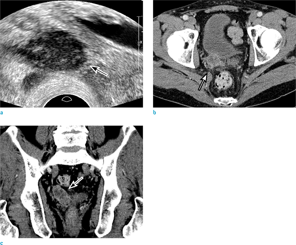
Fig. 1 Primary diffuse large B-cell lymphoma in the seminal vesicle in a 58-year-old man. Transrectal ultrasonography (TRUS) demonstrating a heterogeneous, hypoechoic mass (arrow) in the right seminal vesicle (a). CT images showing a mass with rim-like enhancement (arrows) in the right seminal vesicle on portal venous phase (b, c).
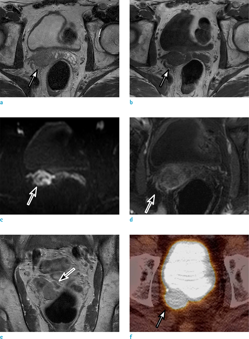
Fig. 2 Primary diffuse large B-cell lymphoma of the seminal vesicle in a 58-year-old man. T2-weighted axial MR image demonstrating a mass with subtle high signal intensity (arrow) in the right seminal vesicle (a); T1-weighted axial image demonstrating a mass of low signal intensity (b, arrow) ; diffusion-weighted imaging showing high intensity (arrow) with a high b-value (1000 s/mm2) (c). The tumor showed rim-like enhancement on 3-min delayed phase image (d, arrow) and progressive with a non-enhancing portion on 5-min delayed phase image (e, arrow). FDG PET/CT revealed a high standardized uptake value (maxSUV, 23.5) in the right seminal vesicle, suggesting a hypermetabolic mass (f, arrow).
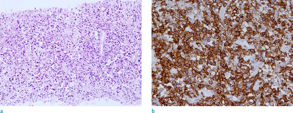
Fig. 3 Primary diffuse large B-cell lymphoma of the seminal vesicle in a 58-year-old man. Staining of the biopsy from the seminal vesicle mass showing diffuse proliferation of large neoplastic lymphoid cells (a: × 200, H&E staining). Immunohistochemical staining showing neoplastic cells expressing CD79a, confirming their B-cell lineage (b: × 400).
Reference
-
1. Kim B, Kawashima A, Ryu JA, Takahashi N, Hartman RP, King BF Jr. Imaging of the seminal vesicle and vas deferens. Radiographics. 2009; 29:1105–1121.2. Dalgaard JB, Giertsen JC. Primary carcinoma of the seminal vesicle; case and survey. Acta Pathol Microbiol Scand. 1956; 39:255–267.3. Benson RC Jr, Clark WR, Farrow GM. Carcinoma of the seminal vesicle. J Urol. 1984; 132:483–485.4. Zhu J, Chen LR, Zhang X, Gong Y, Xu JH, Zheng S. Primary diffuse large B-cell lymphoma of the seminal vesicles: ultrasonography and computed tomography findings. Urology. 2011; 78:1073–1074.5. Zhu B, Cai Y, Chen R, Ye C, Tao Y, Wen X. Primary lymphoma of the seminal vesicles presented with acute renal failure: PET-CT findings. Open J Urol. 2012; 2:137.6. Handa N, Rathinam D, Singh A, Jana M. Seminal vesicle involvement: a rare extranodal manifestation of non-Hodgkin's lymphoma. BMJ Case Rep. 2016; 2016.7. Freeman C, Berg JW, Cutler SJ. Occurrence and prognosis of extranodal lymphomas. Cancer. 1972; 29:252–260.8. Sheth S, Ali S, Fishman E. Imaging of renal lymphoma: patterns of disease with pathologic correlation. Radiographics. 2006; 26:1151–1168.9. Rajiah P, Sinha R, Cuevas C, Dubinsky TJ, Bush WH Jr, Kolokythas O. Imaging of uncommon retroperitoneal masses. Radiographics. 2011; 31:949–976.10. Hamada S, Ito K, Kanbara T, et al. A case of malignant lymphoma mimicking a seminal vesicle tumor. Hinyokika Kiyo. 2010; 56:393–396.
- Full Text Links
-
- Actions
-
Cited
- CITED
-
- Close
- Share
-
- Similar articles
-
- Seminal Vesicle Infection of Zinner Syndrome Misdiagnosed for Neoplasm
- Relapse of Ocular Lymphoma following Primary Testicular Diffuse Large B-cell Lymphoma
- A Case of Primary Cutaneous Diffuse Large B-cell Lymphoma
- Primary Mucinous Adenocarcinoma of a Seminal Vesicle Cyst Associated with Ectopic Ureter and Ipsilateral Renal Agenesis: a Case Report
- Primary Adenocarcinoma of the Seminal Vesicle
