Normal 2-Dimensional Strain Values of the Left Ventricle: A Substudy of the Normal Echocardiographic Measurements in Korean Population Study
- Affiliations
-
- 1Division of Cardiology, Department of Internal Medicine, Chungnam National University Hospital, Chungnam National University School of Medicine, Daejeon, Korea.
- 2Division of Cardiology, Department of Internal Medicine, Chungbuk National University School of Medicine, Cheongju, Korea.
- 3Division of Cardiology, Department of Medicine, Samsung Medical Center, Sungkyunkwan University School of Medicine, Seoul, Korea. parksmc@gmail.com
- 4Division of Cardiology, Department of Internal Medicine, Gil Hospital, Gachon University of Medicine and Science, Incheon, Korea.
- 5Division of Cardiology, Department of Internal Medicine, Incheon St. Mary's Hospital, College of Medicine, The Catholic University of Korea, Incheon, Korea.
- 6Department of Internal Medicine, Seoul St. Mary's Hospital, College of Medicine, The Catholic University of Korea, Seoul, Korea.
- 7Division of Cardiology, Department of Internal Medicine, Gyeongsang National University Hospital, Gyeongsang National University School of Medicine, Jinju, Korea.
- 8Department of Cardiology, Kyung Hee University School of Medicine, Kyung Hee University Hospital at Gangdong, Seoul, Korea.
- 9Division of Cardiology, Keimyung University Dongsan Medical Center, Daegu, Korea.
- 10Division of Cardiology, Department of Internal Medicine, Korea University College of Medicine, Seoul, Korea.
- 11Department of Internal Medicine, Wonkwang University Hospital, Institute of Wonkwang Medical Science, Iksan, Korea.
- 12Division of Cardiology, Department of Internal Medicine, Pusan National University School of Medicine, Busan, Korea.
- 13Division of Cardiology, Department of Internal Medicine, Cardiovascular Center, Seoul National University College of Medicine, Seoul, Korea.
- 14Division of Cardiology, Department of Internal Medicine, Seoul National University and Cardiovascular Center, Seoul National University Bundang Hospital, Seongnam, Korea.
- 15Division of Cardiology, Department of Medicine, Samsung Changwon Hospital, Sungkyunkwan University School of Medicine, Changwon, Korea.
- 16Department of Cardiology, Ajou University School of Medicine, Suwon, Korea.
- 17Division of Cardiology, Severance Cardiovascular Hospital, Yonsei University College of Medicine, Seoul, Korea.
- 18Department of Cardiology, Asan Medical Center, University of Ulsan College of Medicine, Seoul, Korea.
- 19Division of Cardiology, Gangneung Asan Hospital, University of Ulsan College of Medicine, Gangneung, Korea.
- 20Division of Cardiology, Department of Internal Medicine, Ewha Womans University School of Medicine, Seoul, Korea.
- 21Division of Cardiology, Department of Internal Medicine, Inha University College of Medicine, Incheon, Korea.
- 22Department of Cardiology, Chonnam National University Hospital, Gwangju, Korea.
- 23Department of Internal Medicine, Cardiovascular Center, Kyung Hee University Medical Center, Seoul, Korea.
- KMID: 2364646
- DOI: http://doi.org/10.4250/jcu.2016.24.4.285
Abstract
- BACKGROUND
It is important to understand the distribution of 2-dimensional strain values in normal population. We performed a multicenter trial to measure normal echocardiographic values in the Korean population.
METHODS
This was a substudy of the Normal echOcardiogRaphic Measurements in KoreAn popuLation (NORMAL) study. Echocardiographic specialists measured frequently used echocardiographic indices in healthy people according to a standardized method at 23 different university hospitals. The strain values were analyzed from digitally stored images.
RESULTS
Of a total of 1003 healthy participants in NORMAL study, 2-dimensional strain values were measured in 501 subjects (265 females, mean age 47 ± 15 years old) with echocardiographic images only by GE echocardiographic machines. Interventricular septal thickness, left ventricular (LV) posterior wall thickness, systolic and diastolic LV dimensions, and LV ejection fraction were 7.5 ± 1.0 mm, 7.4 ± 1.0 mm, 29.9 ± 2.8 mm, 48.9 ± 3.6 mm, and 62 ± 4%, respectively. LV longitudinal systolic strain (LS) values of apical 4-chamber (A4C) view, apical 3-chamber (A3C) view, apical 2-chamber (A2C) view, and LV global LS (LVGLS) were −20.1 ± 2.3, −19.9 ± 2.7, −21.2 ± 2.6, and −20.4 ± 2.2%, respectively. LV longitudinal systolic strain rate (LVLSR) values of the A4C view, A3C view, A2C view, and LV global LSR (LVGLSR) were −1.18 ± 0.18, −1.20 ± 0.21, −1.25 ± 0.21, and −1.21 ± 0.21(−s), respectively. Females had lower LVGLS (−21.2 ± 2.2% vs. −19.5 ± 1.9%, p < 0.001) and LVGLSR (−1.25 ± 0.18(−s) vs. −1.17 ± 0.15(−s), p < 0.001) values than males.
CONCLUSION
We measured LV longitudinal strain and strain rate values in the normal Korean population. Since considerable gender differences were observed, normal echocardiographic cutoff values should be differentially applied based on sex.
Keyword
MeSH Terms
Figure
-
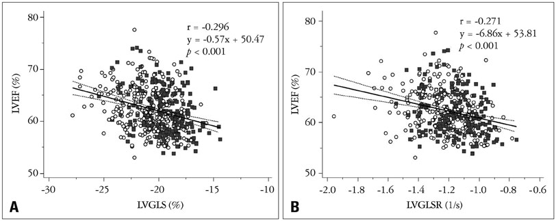
Fig. 1 Correlations between LVEF and LVGLS (A) and LVEF and LVGLSR (B) (open circle: female, filled quadrangle: male). LVEF: left ventricular ejection fraction, LVGLS: left ventricular global longitudinal strain, LVGLSR: left ventricular global longitudinal systolic strain rate.
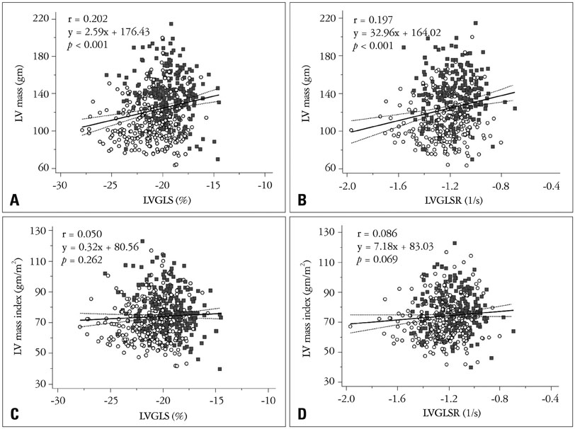
Fig. 2 Correlation between LV mass and LV longitudinal systolic strain. LVGLS and LVGLSR show significant correlations with LV mass (A and B). However, LVGLS and LVGLSR were not significantly correlated with LV mass index (C and D) (open circle: female, filled quadrangle: male). LV: left ventricular, LVGLS: LV global longitudinal strain, LVGLSR: LV global longitudinal systolic strain rate.
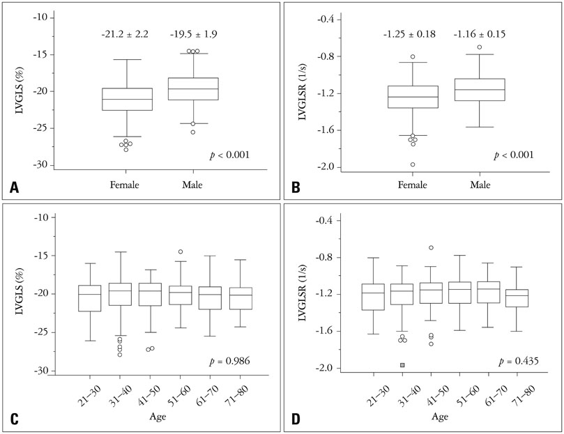
Fig. 3 Females have significantly lower (better) LVGLS (A) and LVGLSR (B) values than those of males. No significant differences were observed among the different age groups (C and D). LVGLS: left ventricular global longitudinal strain, LVGLSR: left ventricular global longitudinal systolic strain rate.
Cited by 3 articles
-
Two-dimensional Echocardiographic Assessment of Myocardial Strain: Important Echocardiographic Parameter Readily Useful in Clinical Field
Jae-Hyeong Park
Korean Circ J. 2019;49(10):908-931. doi: 10.4070/kcj.2019.0200.Time Course of Functional Recovery in Takotsubo (Stress) Cardiomyopathy: A Serial Speckle Tracking Echocardiography and Electrocardiography Study
Mirae Lee
J Cardiovasc Imaging. 2020;28(1):50-60. doi: 10.4250/jcvi.2019.0083.Longitudinal Change in Myocardial Function and Clinical Parameters in Middle-Aged Subjects: A 3-Year Follow-up Study
Dong-Hyuk Cho, Hyung Joon Joo, Mi-Na Kim, Hee-Dong Kim, Do-Sun Lim, Seong-Mi Park
Diabetes Metab J. 2021;45(5):719-729. doi: 10.4093/dmj.2020.0132.
Reference
-
1. Wolk MJ, Bailey SR, Doherty JU, Douglas PS, Hendel RC, Kramer CM, Min JK, Patel MR, Rosenbaum L, Shaw LJ, Stainback RF, Allen JM. American College of Cardiology Foundation Appropriate Use Criteria Task Force. ACCF/AHA/ASE/ASNC/HFSA/HRS/SCAI/SCCT/SCMR/STS 2013 multimodality appropriate use criteria for the detection and risk assessment of stable ischemic heart disease: a report of the American College of Cardiology Foundation Appropriate Use Criteria Task Force, American Heart Association, American Society of Echocardiography, American Society of Nuclear Cardiology, Heart Failure Society of America, Heart Rhythm Society, Society for Cardiovascular Angiography and Interventions, Society of Cardiovascular Computed Tomography, Society for Cardiovascular Magnetic Resonance, and Society of Thoracic Surgeons. J Am Coll Cardiol. 2014; 63:380–406.2. Joyce E, Hoogslag GE, Leong DP, Debonnaire P, Katsanos S, Boden H, Schalij MJ, Marsan NA, Bax JJ, Delgado V. Association between left ventricular global longitudinal strain and adverse left ventricular dilatation after ST-segment-elevation myocardial infarction. Circ Cardiovasc Imaging. 2014; 7:74–81.3. White HD, Norris RM, Brown MA, Takayama M, Maslowski A, Bass NM, Ormiston JA, Whitlock T. Effect of intravenous streptokinase on left ventricular function and early survival after acute myocardial infarction. N Engl J Med. 1987; 317:850–855.4. White HD, Norris RM, Brown MA, Brandt PW, Whitlock RM, Wild CJ. Left ventricular end-systolic volume as the major determinant of survival after recovery from myocardial infarction. Circulation. 1987; 76:44–51.5. Hoit BD. Strain and strain rate echocardiography and coronary artery disease. Circ Cardiovasc Imaging. 2011; 4:179–190.6. Leitman M, Lysyansky P, Sidenko S, Shir V, Peleg E, Binenbaum M, Kaluski E, Krakover R, Vered Z. Two-dimensional strain-a novel software for real-time quantitative echocardiographic assessment of myocardial function. J Am Soc Echocardiogr. 2004; 17:1021–1029.7. Behar V, Adam D, Lysyansky P, Friedman Z. The combined effect of nonlinear filtration and window size on the accuracy of tissue displacement estimation using detected echo signals. Ultrasonics. 2004; 41:743–753.8. Sarvari SI, Haugaa KH, Anfinsen OG, Leren TP, Smiseth OA, Kongsgaard E, Amlie JP, Edvardsen T. Right ventricular mechanical dispersion is related to malignant arrhythmias: a study of patients with arrhythmogenic right ventricular cardiomyopathy and subclinical right ventricular dysfunction. Eur Heart J. 2011; 32:1089–1096.9. Thomas JD, Popović ZB. Assessment of left ventricular function by cardiac ultrasound. J Am Coll Cardiol. 2006; 48:2012–2025.10. Geyer H, Caracciolo G, Abe H, Wilansky S, Carerj S, Gentile F, Nesser HJ, Khandheria B, Narula J, Sengupta PP. Assessment of myocardial mechanics using speckle tracking echocardiography: fundamentals and clinical applications. J Am Soc Echocardiogr. 2010; 23:351–369. quiz 453-5.11. Nahum J, Bensaid A, Dussault C, Macron L, Clémence D, Bouhemad B, Monin JL, Rande JL, Gueret P, Lim P. Impact of longitudinal myocardial deformation on the prognosis of chronic heart failure patients. Circ Cardiovasc Imaging. 2010; 3:249–256.12. Kusunose K, Popović ZB, Motoki H, Marwick TH. Prognostic significance of exercise-induced right ventricular dysfunction in asymptomatic degenerative mitral regurgitation. Circ Cardiovasc Imaging. 2013; 6:167–176.13. Choi JO, Shin MS, Kim MJ, Jung HO, Park JR, Sohn IS, Kim H, Park SM, Yoo NJ, Choi JH, Kim HK, Cho GY, Lee MR, Park JS, Shim CY, Kim DH, Shin DH, Shin GJ, Shin SH, Kim KH, Park JH, Lee SY, Kim WS, Park SW. Normal echocardiographic measurements in a Korean population study: part I. Cardiac chamber and great artery evaluation. J Cardiovasc Ultrasound. 2015; 23:158–172.14. Choi JO, Shin MS, Kim MJ, Jung HO, Park JR, Sohn IS, Kim H, Park SM, Yoo NJ, Choi JH, Kim HK, Cho GY, Lee MR, Park JS, Shim CY, Kim DH, Shin DH, Shin GJ, Shin SH, Kim KH, Park JH, Lee SY, Kim WS, Park SW. Normal echocardiographic measurements in a Korean population study: part II. Doppler and tissue Doppler imaging. J Cardiovasc Ultrasound. 2016; 24:144–152.15. Lang RM, Badano LP, Mor-Avi V, Afilalo J, Armstrong A, Ernande L, Flachskampf FA, Foster E, Goldstein SA, Kuznetsova T, Lancellotti P, Muraru D, Picard MH, Rietzschel ER, Rudski L, Spencer KT, Tsang W, Voigt JU. Recommendations for cardiac chamber quantification by echocardiography in adults: an update from the American Society of Echocardiography and the European Association of Cardiovascular Imaging. J Am Soc Echocardiogr. 2015; 28:1–39.e14.16. Smiseth OA, Torp H, Opdahl A, Haugaa KH, Urheim S. Myocardial strain imaging: how useful is it in clinical decision making? Eur Heart J. 2016; 37:1196–1207.17. Kadappu KK, Thomas L. Tissue Doppler imaging in echocardiography: value and limitations. Heart Lung Circ. 2015; 24:224–233.18. Greenberg NL, Firstenberg MS, Castro PL, Main M, Travaglini A, Odabashian JA, Drinko JK, Rodriguez LL, Thomas JD, Garcia MJ. Doppler-derived myocardial systolic strain rate is a strong index of left ventricular contractility. Circulation. 2002; 105:99–105.19. Park JH, Negishi K, Kwon DH, Popovic ZB, Grimm RA, Marwick TH. Validation of global longitudinal strain and strain rate as reliable markers of right ventricular dysfunction: comparison with cardiac magnetic resonance and outcome. J Cardiovasc Ultrasound. 2014; 22:113–120.20. Kovács A, Oláh A, Lux Á, Mátyás C, Németh BT, Kellermayer D, Ruppert M, Török M, Szabó L, Meltzer A, Assabiny A, Birtalan E, Merkely B, Radovits T. Strain and strain rate by speckle-tracking echocardiography correlate with pressure-volume loop-derived contractility indices in a rat model of athlete’s heart. Am J Physiol Heart Circ Physiol. 2015; 308:H743–H748.21. Yingchoncharoen T, Agarwal S, Popović ZB, Marwick TH. Normal ranges of left ventricular strain: a meta-analysis. J Am Soc Echocardiogr. 2013; 26:185–191.22. Takigiku K, Takeuchi M, Izumi C, Yuda S, Sakata K, Ohte N, Tanabe K, Nakatani S. JUSTICE investigators. Normal range of left ventricular 2-dimensional strain: Japanese Ultrasound Speckle Tracking of the Left Ventricle (JUSTICE) study. Circ J. 2012; 76:2623–2632.23. Kocabay G, Muraru D, Peluso D, Cucchini U, Mihaila S, Padayattil-Jose S, Gentian D, Iliceto S, Vinereanu D, Badano LP. Normal left ventricular mechanics by two-dimensional speckle-tracking echocardiography. Reference values in healthy adults. Rev Esp Cardiol (Engl Ed). 2014; 67:651–658.24. Saito K, Okura H, Watanabe N, Hayashida A, Obase K, Imai K, Maehama T, Kawamoto T, Neishi Y, Yoshida K. Comprehensive evaluation of left ventricular strain using speckle tracking echocardiography in normal adults: comparison of three-dimensional and two-dimensional approaches. J Am Soc Echocardiogr. 2009; 22:1025–1030.25. Caselli S, Montesanti D, Autore C, Di Paolo FM, Pisicchio C, Squeo MR, Musumeci B, Spataro A, Pandian NG, Pelliccia A. Patterns of left ventricular longitudinal strain and strain rate in Olympic athletes. J Am Soc Echocardiogr. 2015; 28:245–253.26. Dalen H, Thorstensen A, Aase SA, Ingul CB, Torp H, Vatten LJ, Stoylen A. Segmental and global longitudinal strain and strain rate based on echocardiography of 1266 healthy individuals: the HUNT study in Norway. Eur J Echocardiogr. 2010; 11:176–183.27. Andre F, Steen H, Matheis P, Westkott M, Breuninger K, Sander Y, Kammerer R, Galuschky C, Giannitsis E, Korosoglou G, Katus HA, Buss SJ. Age- and gender-related normal left ventricular deformation assessed by cardiovascular magnetic resonance feature tracking. J Cardiovasc Magn Reson. 2015; 17:25.28. Staron A, Bansal M, Kalakoti P, Nakabo A, Gasior Z, Pysz P, Wita K, Jasinski M, Sengupta PP. Speckle tracking echocardiography derived 2-dimensional myocardial strain predicts left ventricular function and mass regression in aortic stenosis patients undergoing aortic valve replacement. Int J Cardiovasc Imaging. 2013; 29:797–808.
- Full Text Links
-
- Actions
-
Cited
- CITED
-
- Close
- Share
-
- Similar articles
-
- Two-dimensional Echocardiographic Assessment of Myocardial Strain: Important Echocardiographic Parameter Readily Useful in Clinical Field
- Multicenter Trial for Estimation of Normal Values of Echocardiographic Indices in Korea
- Strain Analysis of the Right Ventricle Using Two-dimensional Echocardiography
- Systolic Long Axis Function of the Left Ventricle, as Assessed by 2-D Strain, is Reduced in the Patients Who Have Diastolic Dysfunction and a Normal Ejection Fraction
- Tissue Doppler and strain imaging of left ventricle in Beagle dogs with iatrogenic hypercortisolism
