Imaging Sci Dent.
2016 Dec;46(4):245-249. 10.5624/isd.2016.46.4.245.
Analysis of the priority of anatomic structures according to the diagnostic task in cone-beam computed tomographic images
- Affiliations
-
- 1Department of Oral and Maxillofacial Radiology, Dankook University College of Dentistry, Chungnam, Korea. runnachv@dankook.ac.kr
- KMID: 2362742
- DOI: http://doi.org/10.5624/isd.2016.46.4.245
Abstract
- PURPOSE
This study was designed to evaluate differences in the required visibility of anatomic structures according to the diagnostic tasks of implant planning and periapical diagnosis.
MATERIALS AND METHODS
Images of a real skull phantom were acquired under 24 combinations of different exposure conditions in a cone-beam computed tomography scanner (60, 70, 80, 90, 100, and 110 kV and 4, 6, 8, and 10 mA). Five radiologists evaluated the visibility of anatomic structures and the image quality for diagnostic tasks using a 6-point scale.
RESULTS
The visibility of the periodontal ligament space showed the closest association with the ability to use an image for periapical diagnosis in both jaws. The visibility of the sinus floor and canal wall showed the closest association with the ability to use an image for implant planning. Variations in tube voltage were associated with significant differences in image quality for all diagnostic tasks. However, tube current did not show significant associations with the ability to use an image for implant planning.
CONCLUSION
The required visibility of anatomic structures varied depending on the diagnostic task. Tube voltage was a more important exposure parameter for image quality than tube current. Different settings should be used for optimization and image quality evaluation depending on the diagnostic task.
Figure
-
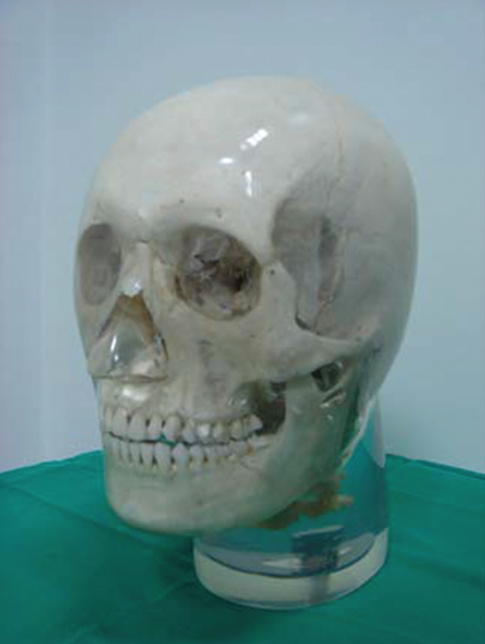
Fig. 1 Transparent X-ray skull phantom with a soft tissue replica.
Cited by 2 articles
-
Factors affecting modulation transfer function measurements in cone-beam computed tomographic images
Jin-Woo Choi
Imaging Sci Dent. 2019;49(2):131-137. doi: 10.5624/isd.2019.49.2.131.Optimization of exposure parameters and relationship between subjective and technical image quality in cone-beam computed tomography
Ha-Na Park, Chang-Ki Min, Kyoung-A Kim, Kwang-Joon Koh
Imaging Sci Dent. 2019;49(2):139-151. doi: 10.5624/isd.2019.49.2.139.
Reference
-
1. Ludlow JB, Ivanovic M. Comparative dosimetry of dental CBCT devices and 64-slice CT for oral and maxillofacial radiology. Oral Surg Oral Med Oral Pathol Oral Radiol Endod. 2008; 106:106–114.
Article2. Goulston R, Davies J, Horner K, Murphy F. Dose optimization by altering the operating potential and tube current exposure time product in dental cone beam CT: a systematic review. Dentomaxillofac Radiol. 2016; 45:20150254.
Article3. Pauwels R, Seynaeve L, Henriques JC, de Oliveira-Santos C, Souza PC, Westphalen FH, et al. Optimization of dental CBCT exposures through mAs reduction. Dentomaxillofac Radiol. 2015; 44:20150108.
Article4. Pauwels R, Beinsberger J, Stamatakis H, Tsiklakis K, Walker A, Bosmans H, et al. Comparison of spatial and contrast resolution for cone-beam computed tomography scanners. Oral Surg Oral Med Oral Pathol Oral Radiol. 2012; 114:127–135.
Article5. Bamba J, Araki K, Endo A, Okano T. Image quality assessment of three cone beam CT machines using the SEDENTEXCT CT phantom. Dentomaxillofac Radiol. 2013; 42:20120445.
Article6. Watanabe H, Honda E, Kurabayashi T. Modulation transfer function evaluation of cone beam computed tomography for dental use with the oversampling method. Dentomaxillofac Radiol. 2010; 39:28–32.
Article7. Xu J, Reh DD, Carey JP, Mahesh M, Siewerdsen JH. Technical assessment of a cone-beam CT scanner for otolaryngology imaging: image quality, dose, and technique protocols. Med Phys. 2012; 39:4932–4942.
Article8. Ozaki Y, Watanabe H, Nomura Y, Honda E, Sumi Y, Kurabayashi T. Location dependency of the spatial resolution of cone beam computed tomography for dental use. Oral Surg Oral Med Oral Pathol Oral Radiol. 2013; 116:648–655.
Article9. Lofthag-Hansen S, Thilander-Klang A, Grondahl K. Evaluation of subjective image quality in relation to diagnostic task for cone beam computed tomography with different fields of view. Eur J Radiol. 2011; 80:483–488.
Article10. Tapiovaara M. Review of relationships between physical measurements and user evaluation of image quality. Radiat Prot Dosimetry. 2008; 129:244–248.
Article11. Choi JW, Lee SS, Choi SC, Heo MS, Huh KH, Yi WJ, et al. Relationship between physical factors and subjective image quality of cone-beam computed tomography images according to diagnostic task. Oral Surg Oral Med Oral Pathol Oral Radiol. 2015; 119:357–365.
Article12. De Cock J, Zanca F, Canning J, Pauwels R, Hermans R. A comparative study for image quality and radiation dose of a cone beam computed tomography scanner and a multislice computed tomography scanner for paranasal sinus imaging. Eur Radiol. 2015; 25:1891–1900.
Article13. Dawood A, Brown J, Sauret-Jackson V, Purkayastha S. Optimization of cone beam CT exposure for pre-surgical evaluation of the implant site. Dentomaxillofac Radiol. 2012; 41:70–74.
Article14. Sur J, Seki K, Koizumi H, Nakajima K, Okano T. Effects of tube current on cone-beam computerized tomography image quality for presurgical implant planning in vitro. Oral Surg Oral Med Oral Pathol Oral Radiol Endod. 2010; 110:e29–e33.
Article15. Liang X, Jacobs R, Hassan B, Li L, Pauwels R, Corpas L, et al. A comparative evaluation of Cone Beam Computed Tomography (CBCT) and Multi-Slice CT (MSCT) Part I. On subjective image quality. Eur J Radiol. 2010; 75:265–269.
- Full Text Links
-
- Actions
-
Cited
- CITED
-
- Close
- Share
-
- Similar articles
-
- The impact of reorienting cone-beam computed tomographic images in varied head positions on the coordinates of anatomical landmarks
- Cone beam CT findings of retromolar canals: Report of cases and literature review
- Mandibular canal branches supplying the mandibular third molar observed on cone beam computed tomographic images: Reports of four cases
- Cone beam computed tomography findings of ectopic mandibular third molar in the mandibular condyle: report of a case
- Ameloblastoma with dystrophic calcification: A case report with 3-dimensional cone-beam computed tomographic images of calcification
