Imaging Sci Dent.
2016 Jun;46(2):133-139. 10.5624/isd.2016.46.2.133.
The impact of reorienting cone-beam computed tomographic images in varied head positions on the coordinates of anatomical landmarks
- Affiliations
-
- 1Department of Oral and Maxillofacial Radiology, Yonsei University, College of Dentistry, Seoul, Korea. sshan@yuhs.ac rari98@yuhs.ac
- KMID: 2308877
- DOI: http://doi.org/10.5624/isd.2016.46.2.133
Abstract
- PURPOSE
The aim of this study was to compare the coordinates of anatomical landmarks on cone-beam computed tomographic (CBCT) images in varied head positions before and after reorientation using image analysis software.
MATERIALS AND METHODS
CBCT images were taken in a normal position and four varied head positions using a dry skull marked with 3 points where gutta percha was fixed. In each of the five radiographic images, reference points were set, 20 anatomical landmarks were identified, and each set of coordinates was calculated. Coordinates in the images from the normally positioned head were compared with those in the images obtained from varied head positions using statistical methods. Post-reorientation coordinates calculated using a three-dimensional image analysis program were also compared to the reference coordinates.
RESULTS
In the original images, statistically significant differences were found between coordinates in the normal-position and varied-position images. However, post-reorientation, no statistically significant differences were found between coordinates in the normal-position and varied-position images.
CONCLUSION
The changes in head position impacted the coordinates of the anatomical landmarks in three-dimensional images. However, reorientation using image analysis software allowed accurate superimposition onto the reference positions.
MeSH Terms
Figure
-
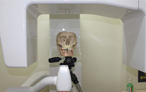
Fig. 1 A dry skull fixed to the tripod for cone-beam computed tomography.
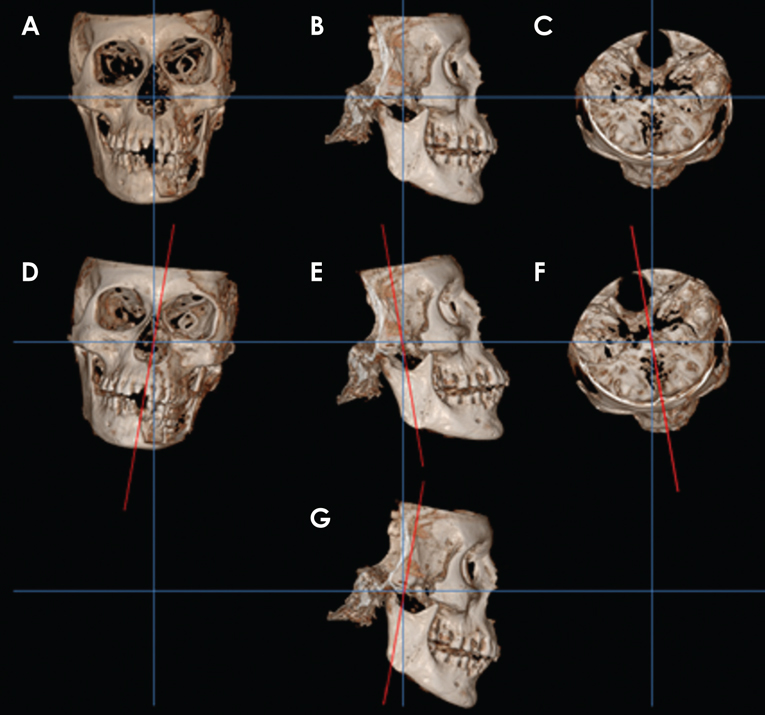
Fig. 2 Three-dimensional cone-beam computed tomography images corresponding to 5 head positions. A. Normal position (horizontal plane). B. Normal position (sagittal plane). C. Normal position (axial plane). D. Five-degree leftward tilting. E. Five-degree extension. F. Five-degree leftward rotation. G. Five-degree flexion.
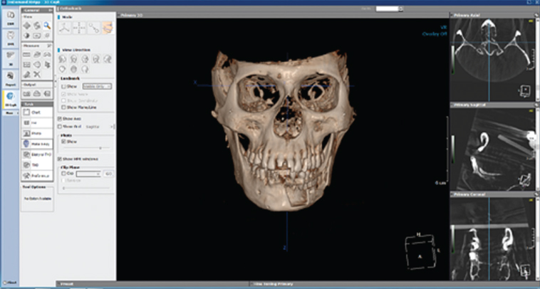
Fig. 3 Reconstructed three-dimensional cone-beam computed tomography images by OnDemand 3D™.
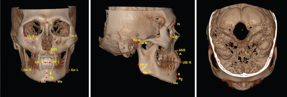
Fig. 4 Twenty landmarks are seen on the reconstructed three-dimensional image.
Reference
-
1. El-Beialy AR, Fayed MS, El-Bialy AM, Mostafa YA. Accuracy and reliability of cone-beam computed tomography measurements: influence of head orientation. Am J Orthod Dentofacial Orthop. 2011; 140:157–165.
Article2. Kau C, Richmond S, Palomo J, Hans M. Three-dimensional cone beam computerized tomography in orthodontics. J Orthod. 2005; 32:282–293.3. Sheikhi M, Ghorbanizadeh S, Abdinian M, Goroohi H, Badrian H. Accuracy of linear measurements of Galileos cone beam computed tomography in normal and different head positions. Int J Dent. 2012; 2012:214954.
Article4. van Steenberghe D, Naert I, Andersson M, Brajnovic I, Van Cleynenbreugel J, Suetens P. A custom template and definitive prosthesis allowing immediate implant loading in the maxilla: a clinical report. Int J Oral Maxillofac Implants. 2002; 17:663–670.5. Gahleitner A, Watzek G, Imhof H. Dental CT: imaging technique, anatomy, and pathologic conditions of the jaws. Eur Radiol. 2003; 13:366–376.
Article6. Cohnen M, Kemper J, Möbes O, Pawelzik J, Mödder U. Radiation dose in dental radiology. Eur Radiol. 2002; 12:634–637.
Article7. Hein E, Rogalla P, Klingebiel R, Hamm B. Low-dose CT of the paranasal sinuses with eye lens protection: effect on image quality and radiation dose. Eur Radiol. 2002; 12:1693–1696.
Article8. Hagtvedt T, Aaløkken TM, Nøtthellen J, Kolbenstvedt A. A new low-dose CT examination compared with standard-dose CT in the diagnosis of acute sinusitis. Eur Radiol. 2003; 13:976–980.
Article9. Mah JK, Danforth RA, Bumann A, Hatcher D. Radiation absorbed in maxillofacial imaging with a new dental computed tomography device. Oral Surg Oral Med Oral Pathol Oral Radiol Endod. 2003; 96:508–513.
Article10. Ludlow JB, Davies-Ludlow LE, Brooks SL. Dosimetry of two extraoral direct digital imaging devices: NewTom cone beam CT and Orthophos Plus DS panoramic unit. Dentomaxillofac Radiol. 2003; 32:229–234.
Article11. Sukovic P. Cone beam computed tomography in craniofacial imaging. Orthod Craniofac Res. 2003; 6:Suppl 1. 31–36.
Article12. Hashimoto K, Arai Y, Iwai K, Araki M, Kawashima S, Terakado M. A comparison of a new limited cone beam computed tomography machine for dental use with a multidetector row helical CT machine. Oral Surg Oral Med Oral Pathol Oral Radiol Endod. 2003; 95:371–377.
Article13. Ziegler CM, Woertche R, Brief J, Hassfeld S. Clinical indications for digital volume tomography in oral and maxillofacial surgery. Dentomaxillofac Radiol. 2002; 31:126–130.
Article14. Hassan B, van der Stelt P, Sanderink G. Accuracy of three-dimensional measurements obtained from cone beam computed tomography surface-rendered images for cephalometric analysis: influence of patient scanning position. Eur J Orthod. 2009; 31:129–134.
Article15. Togashi K, Kitaura H, Yonetsu K, Yoshida N, Nakamura T. Three-dimensional cephalometry using helical computer tomography: measurement error caused by head inclination. Angle Orthod. 2002; 72:513–520.16. Lagravere MO, Major PW, Carey J. Sensitivity analysis for plane orientation in three-dimensional cephalometric analysis based on superimposition of serial cone beam computed tomography images. Dentomaxillofac Radiol. 2010; 39:400–408.17. Kitaura H, Yonetsu K, Kitamori H, Kobayashi K, Nakamura T. Standardization of 3-D CT measurements for length and angles by matrix transformation in the 3-D coordinate system. Cleft Palate Craniofac J. 2000; 37:349–356.
Article18. Hwang JJ, Kim KD, Park H, Park CS, Jeong HG. Factors influencing superimposition error of 3D cephalometric landmarks by plane orientation method using 4 reference points: 4 point superimposition error regression model. PLoS One. 2014; 9:e110665.
Article19. Sabban H, Mahdian M, Dhingra A, Lurie AG, Tadinada A. Evaluation of linear measurements of implant sites based on head orientation during acquisition: an ex vivo study using cone-beam computed tomography. Imaging Sci Dent. 2015; 45:73–80.
Article20. de Oliveira AE, Cevidanes LH, Phillips C, Motta A, Burke B, Tyndall D. Observer reliability of three-dimensional cephalometric landmark identification on cone-beam computerized tomography. Oral Surg Oral Med Oral Pathol Oral Radiol Endod. 2009; 107:256–265.
Article21. Berco M, Rigali PH Jr, Miner RM, DeLuca S, Anderson NK, Will LA. Accuracy and reliability of linear cephalometric measurements from cone-beam computed tomography scans of a dry human skull. Am J Orthod Dentofacial Orthop. 2009; 136:17.e1–17.e9.
Article22. Ludlow JB, Gubler M, Cevidanes L, Mol A. Precision of cephalometric landmark identification: cone-beam computed tomography vs conventional cephalometric views. Am J Orthod Dentofacial Orthop. 2009; 136:312.e1–312.e10.
Article
- Full Text Links
-
- Actions
-
Cited
- CITED
-
- Close
- Share
-
- Similar articles
-
- Effect of Voxel Size on the Accuracy of Landmark Identification in Cone-Beam Computed Tomography Images
- Morphometric analysis of the inter-mastoid triangle for sex determination: Application of statistical shape analysis
- A method for mandibular dental arch superimposition using 3D cone beam CT and orthodontic 3D digital model
- Linear accuracy of cone-beam computed tomography and a 3-dimensional facial scanning system: An anthropomorphic phantom study
- Cone beam CT findings of retromolar canals: Report of cases and literature review
