Corneal Collagen Crosslinking in Progressive Keratoconus
- Affiliations
-
- 1Department of Ophthalmology and Visual Science, College of Medicine, The Catholic University of Korea, Seoul, Korea. ckjoo@catholic.ac.kr
- KMID: 2357726
- DOI: http://doi.org/10.3341/jkos.2016.57.11.1714
Abstract
- PURPOSE
To report the clinical efficacy and safety of progressive keratoconic eyes in Korean patients treated with accelerated corneal cross-linking.
METHODS
This retrospective study focused on progressive keratoconic eyes in Korean patients that underwent accelerated corneal cross-linking from February 2015 to October 2015. Keratoconus was diagnosed in 45 eyes in 30 patients. After accelerated corneal cross-linking with VibeX rapid solution, best corrected visual acuity, maximum keratometry, mean keratometry, corneal thickness, corneal astigmatism, and endothelial cell count were measured at the preoperative visit and post operation 1 week, 1 month, 3 months, and 6 months.
RESULTS
Best corrected visual acuity (log MAR) was 0.51 ± 0.23 at pre operation and 0.51 ± 0.26 at post operation 6 months, showing no improvement. The maximum keratometry measured with Auto K, Pentacam, and Orbscan II at pre operation was 49.11 ± 4.5 D, 48.37 ± 3.31 D, and 48.98 ± 4.88 D and changed to 49.29 ± 4.34 D, 46.99 ± 3.63 D, and 47.01 ± 3.62 D postoperatively, respectively. Only Pentacam and Orbscan II measurements showed a statistically significant decrease (p < 0.05). Corneal thickness (at the thinnest area) was measured with Pentacam and Orbscan II; pre-operative and post-operative 6 month data showed changes from 485 ± 26.27 and 479.24 ± 27.89 to 471.64 ± 27.12 and 472.52 ± 25.36, respectively. Only the Pentacam method resulted in a statistically significant decrease. Endothelial cell count was measured with confocal microscopy and showed a statistically significant difference between pre-operative 2,857 ± 390.49/mm² and post-operative 6 month 2,639.21 ± 249.92/mm².
CONCLUSIONS
This 6-month follow-up study of Korean keratoconus patients who underwent accelerated corneal cross-linking indicates that the method is effective in stabilizing the progression of keratoconus, according to maximum keratometry change. With regard to endothelial cell count change, further long-term evaluation is required. Other than endothelial cell count change, this procedure is expected to show long-term safety comparable to that of conventional corneal cross-linking.
MeSH Terms
Figure
-
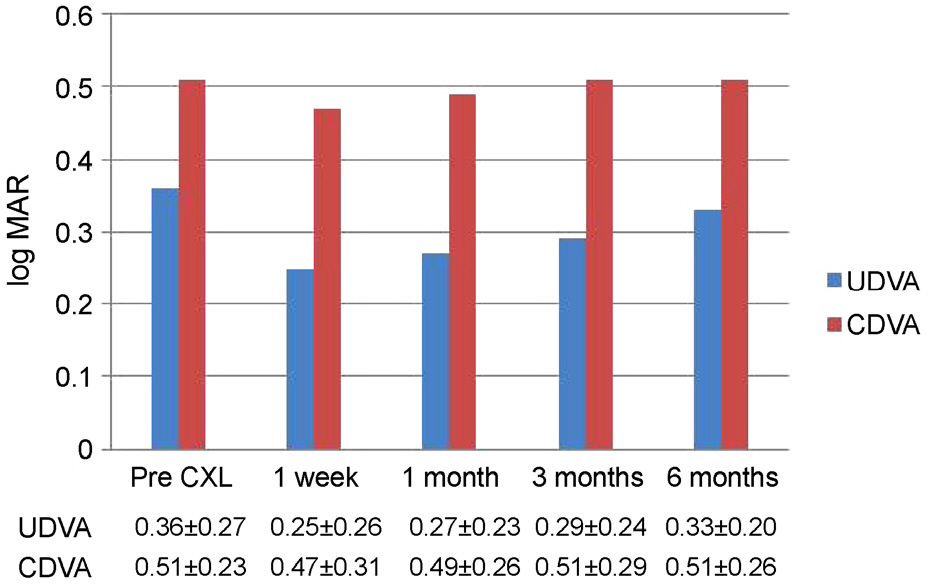
Figure 1. Visual acuity changes in patients. The mean log MAR ± standard deviation, uncorrected and corrected distance visual acuity of patients with keratoconus with thin corneas before and after the accelerated corneal collagen cross-linking. CXL = corneal collagen cross-linking; UDVA = uncorrected distance visual acuity; CDVA = corrected distance visual acuity.

Figure 2. Keratometric changes in patients. The mean ± standard deviation of keratometric changes in Auto Kmax, Pentacam Kmax, ORB scan Kmax before and 1 week, 1 month, 3 month, and 6 months after the accelerated corneal collagen crosslinking in patients with keratoconus with thin corneas. CXL = corneal collagen cross-linking.
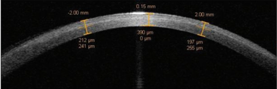
Figure 3. Image of demarcation line. Corneal stromal demarcation line image with anterior segment optical coherence tomography 1 month after accelerated corneal collagen cross-linking ([CXL] 8 minutes at 30 mW/cm2, total surface dose of 7.2 J/cm2).
Reference
-
References
1. Rabinowitz YS. Keratoconus. Surv Ophthalmol. 1998; 42:297–319.
Article2. Wollensak G, Spoerl E, Seiler T. Riboflavin/ultraviolet-a-induced collagen crosslinking for the treatment of keratoconus. Am J Ophthalmol. 2003; 135:620–7.
Article3. Caporossi A, Baiocchi S, Mazzotta C, et al. Parasurgical therapy for keratoconus by riboflavin-ultraviolet type A rays induced abdominal of corneal collagen: preliminary refractive results in an Italian study. J Cataract Refract Surg. 2006; 32:837–45.4. Caporossi A, Mazzotta C, Baiocchi S, Caporossi T. abdominal abdominal of riboflavin ultraviolet a corneal collagen abdominal for keratoconus in Italy: the Siena eye cross study. Am J Ophthalmol. 2010; 149:585–93.5. Raiskup-Wolf F, Hoyer A, Spoerl E, Pillunat LE. Collagen abdominal with riboflavin and ultraviolet-A light in keratoconus: long-term results. J Cataract Refract Surg. 2008; 34:796–801.6. Spoerl E, Huhle M, Seiler T. Induction of cross-links in corneal tissue. Exp Eye Res. 1998; 66:97–103.
Article7. Spoerl E, Wollensak G, Seiler T. Increased resistance of cross-linked cornea against enzymatic digestion. Curr Eye Res. 2004; 29:35–40.
Article8. Gore DM, Margineanu A, French P, et al. Two-photon fluorescence microscopy of corneal riboflavin absorption. Invest Ophthalmol Vis Sci. 2014; 55:2476–81.
Article9. Koller T, Mrochen M, Seiler T. Complication and failure rates after corneal crosslinking. J Cataract Refract Surg. 2009; 35:1358–62.
Article10. Rocha KM, Ramos-Esteban JC, Qian Y, et al. Comparative study of riboflavin-UVA abdominal and “flash-linking” using surface wave elastometry. J Refract Surg. 2008; 24:S748–51.
Article11. Bunsen RW, Roscoe HE. Photochemical Researches.–Part V. On the measurement of the chemical action of direct and diffuse sunlight. Proceedings of the Royal Society of London. 1862; 12:306–12.12. Mrochen M. Current status of accelerated corneal abdominal. Indian J Ophthalmol. 2013; 61:428–9.13. Kymionis GD, Tsoulnaras KI, Grentzelos MA, et al. Evaluation of corneal stromal demarcation line depth following standard and a modified-accelerated collagen abdominal protocol. Am J Ophthalmol. 2014; 158:671–5.e1.14. Hammer A, Richoz O, Arba Mosquera S, et al. Corneal abdominal properties at different corneal abdominal (CXL) irradiances. Invest Ophthalmol Vis Sci. 2014; 55:2881–4.15. Lee P, Jin KH. Clinical results of riboflavin and ultraviolet-a-induced corneal abdominal for progressive keratoconus in Korean patients. J Korean Ophthalmol Soc. 2011; 52:23–8.16. Maeda N, Klyce SD, Smolek MK. Comparison of methods for abdominaling keratoconus using videokeratography. Arch Ophthalmol. 1995; 113:870–4.17. Wittig-Silva C, Whiting M, Lamoureux E, et al. A randomized abdominalled trial of corneal collagen abdominal in progressive abdominal: preliminary results. J Refract Surg. 2008; 24:S720–5.
Article18. Agrawal V. abdominal results of cornea collagen abdominal with riboflavin for keratoconus. Indian J Ophthalmol. 2013; 61:433–4.19. Hashemi H, Seyedian MA, Miraftab M, et al. Corneal collagen abdominal with riboflavin and ultraviolet a irradiation for abdominal: long-term results. Ophthalmology. 2013; 120:1515–20.20. Schumacher S, Oeftiger L, Mrochen M. Equivalence of abdominal changes induced by rapid and standard corneal abdominal, using riboflavin and ultraviolet radiation. Invest Ophthalmol Vis Sci. 2011; 52:9048–52.21. Mita M, Waring GO 4th, Tomita M. High-irradiance accelerated collagen crosslinking for the treatment of keratoconus: six-month results. J Cataract Refract Surg. 2014; 40:1032–40.
Article22. Elbaz U, Shen C, Lichtinger A, et al. Accelerated (9-mW/cm2) abdominal collagen crosslinking for keratoconus-A 1-year follow-up. Cornea. 2014; 33:769–73.23. Cinar Y, Cingü AK, Türkcü FM, et al. Comparison of accelerated and conventional corneal collagen abdominal for progressive keratoconus. Cutan Ocul Toxicol. 2014; 33:218–22.24. Vinciguerra P, Albè E, Trazza S, et al. Intraoperative and abdominal effects of corneal collagen abdominal on progressive keratoconus. Arch Ophthalmol. 2009; 127:1258–65.25. Davis LJ, Schechtman KB, Begley CG, et al. Repeatability of abdominal and corrected visual acuity in keratoconus. The CLEK Study Group. Collaborative Longitudinal Evaluation of Keratoconus. Optom Vis Sci. 1998; 75:887–96.26. Raasch TW, Schechtman KB, Davis LJ, Zadnik K. Repeatability of subjective refraction in myopic and keratoconic subjects: results of vector analysis. Ophthalmic Physiol Opt. 2001; 21:376–83.
Article27. Asri D, Touboul D, Fournié P, et al. Corneal collagen crosslinking in progressive keratoconus: multicenter results from the French National Reference Center for Keratoconus. J Cataract Refract Surg. 2011; 37:2137–43.
Article28. Tomita M, Mita M, Huseynova T. Accelerated versus conventional corneal collagen crosslinking. J Cataract Refract Surg. 2014; 40:1013–20.
Article29. Greenstein SA, Shah VP, Fry KL, Hersh PS. Corneal thickness changes after corneal collagen crosslinking for keratoconus and corneal ectasia: one-year results. J Cataract Refract Surg. 2011; 37:691–700.
Article30. Kontadakis GA, Ginis H, Karyotakis N, et al. In vitro effect of abdominal collagen abdominal on corneal hydration properties and stiffness. Graefes Arch Clin Exp Ophthalmol. 2013; 251:543–7.31. Mazzotta C, Caporossi T, Denaro R, et al. Morphological and functional correlations in riboflavin UV A corneal collagen abdominal for keratoconus. Acta Ophthalmol. 2012; 90:259–65.32. Doors M, Tahzib NG, Eggink FA, et al. Use of anterior segment abdominalal coherence tomography to study corneal changes after collagen abdominal. Am J Ophthalmol. 2009; 148:844–51.e2.33. Hafezi F, Kanellopoulos J, Wiltfang R, Seiler T. Corneal collagen crosslinking with riboflavin and ultraviolet A to treat induced abdominal after laser in situ keratomileusis. J Cataract Refract Surg. 2007; 33:2035–40.34. Gokhale NS. Corneal endothelial damage after collagen abdominal treatment. Cornea. 2011; 30:1495–8.35. Niederer RL, Perumal D, Sherwin T, McGhee CN. Laser scanning in vivo confocal microscopy reveals reduced innervation and abdominal in cell density in all layers of the keratoconic cornea. Invest Ophthalmol Vis Sci. 2008; 49:2964–70.36. al-Abdulla NA, Jabbur NS, O'Brien TP. Astigmatism outcomes following spherical photorefractive keratectomy for myopia. J Refract Surg. 1998; 14:610–4.
Article37. Wilson SE, He YG, Weng J, et al. Epithelial injury induces kerato-cyte apoptosis: hypothesized role for the interleukin-1 system in the modulation of corneal tissue organization and wound healing. Exp Eye Res. 1996; 62:325–7.
Article38. Kymionis GD, Portaliou DM, Diakonis VF, et al. Corneal collagen abdominal with riboflavin and ultraviolet-A irradiation in abdominals with thin corneas. Am J Ophthalmol. 2012; 153:24–8.39. Kymionis GD, Diakonis VF, Kalyvianaki M, et al. One-year fol-low-up of corneal confocal microscopy after corneal abdominal in patients with post laser in situ keratosmileusis ectasia and keratoconus. Am J Ophthalmol. 2009; 147:774–8. 778.e1.
- Full Text Links
-
- Actions
-
Cited
- CITED
-
- Close
- Share
-
- Similar articles
-
- Ten-year Results after Conventional Corneal Cross-linking in Korean Patients with Progressive Keratoconus
- Corneal Crosslinking in Far-Advanced Keratoconus
- Effect of Sequential Intrastromal Corneal Ring Segment Implantation and Corneal Collagen Crosslinking in Corneal Ectasia
- Anterior Elevation Changes Following Corneal Crosslinking for Keratoconus
- Changes in Corneal Keratometry Readings after Corneal Collagen Cross-Linking Using Alcohol in Keratoconus Patients
