Korean J Neurotrauma.
2016 Oct;12(2):144-147. 10.13004/kjnt.2016.12.2.144.
A Case of Intracranial Wooden Foreign Body: Mimicking Pneumocephalus
- Affiliations
-
- 1Department of Neurosurgery, Ulsan University Hospital, University of Ulsan College of Medicine, Ulsan, Korea. esparkns@naver.com
- KMID: 2356789
- DOI: http://doi.org/10.13004/kjnt.2016.12.2.144
Abstract
- Intracranial wooden foreign bodies are rare. In addition, such objects are difficult to identify with conventional radiographic techniques, such as X-ray radiography or brain computed tomography. A 48-year-old man presented to our emergency room with a headache. Even though he had a history of trauma, he had no external wounds and showed no neurological deficits at the initial examination. He was initially diagnosed with trauma-related pneumocephalus. He developed a delayed intracranial infection and underwent surgery to remove the wooden foreign body. The present case illustrates the necessity for special attention to patients suspected of having pneumocephalus with a rare presentation during the initial examination. Early surgical removal of the intracranial foreign body is necessary to prevent complications.
Keyword
MeSH Terms
Figure
-
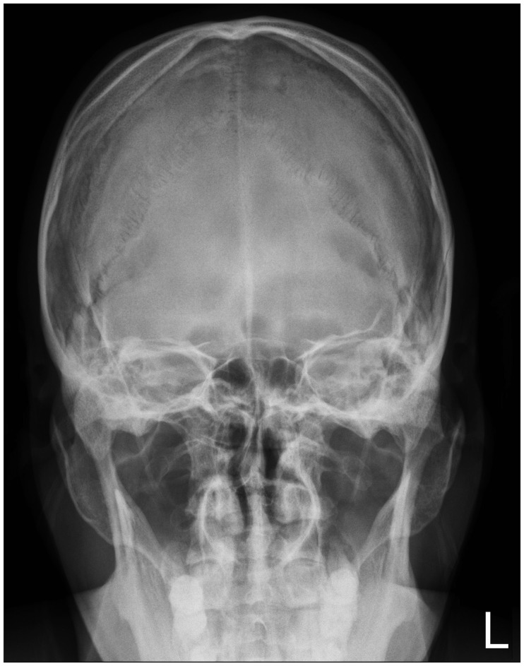
FIGURE 1 Skull X-ray anterior-posterior view showing no abnormal lesions.
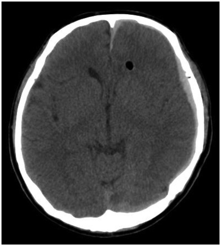
FIGURE 2 A brain computed tomography scan with an axial view demonstrating air density in the left frontal lobe and subdural hematoma on the left convexity.
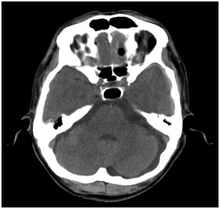
FIGURE 3 A brain computed tomography scan with an axial view demonstrating air density of nasal cavity.
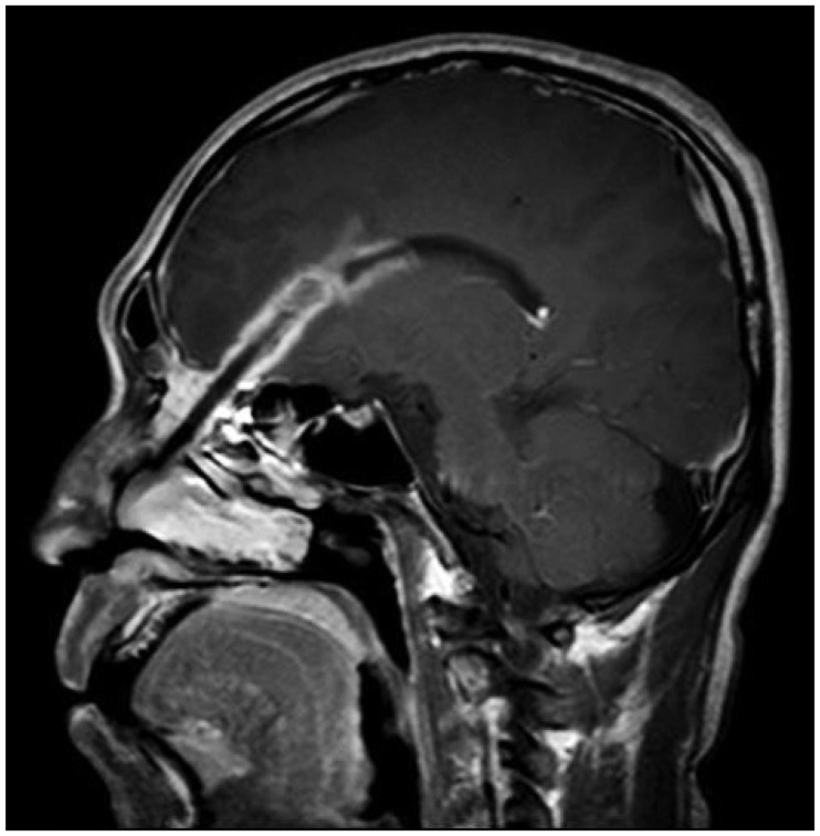
FIGURE 4 T1 gadolinium-enhanced brain magnetic resonance image with a sagittal view showing a track-like intracranial enhancement of the suspected foreign body.
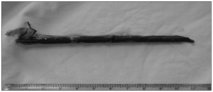
FIGURE 5 Photograph of the wooden foreign object that was removed by a transnasal endoscopic operation.
Reference
-
1. Arifin MZ, Gill AS, Faried A. Penetrating skull fracture by a wooden object: Management dilemmas and literature review. Asian J Neurosurg. 2012; 7:131–134. PMID: 23293668.
Article2. Aulino JM, Gyure KA, Morton A, Cole JW. Temporal lobe intraparenchymal retained foreign body from remote orbital trauma. AJNR Am J Neuroradiol. 2005; 26:1855–1857. PMID: 16091543.3. Dalley RW. Intraorbital wood foreign bodies on CT: use of wide bone window settings to distinguish wood from air. AJR Am J Roentgenol. 1995; 164:434–435. PMID: 7839984.
Article4. Hansen JE, Gudeman SK, Holgate RC, Saunders RA. Penetrating intracranial wood wounds: clinical limitations of computerized tomography. J Neurosurg. 1988; 68:752–756. PMID: 3357035.
Article5. Horner FA, Berry RG, Frantz M. Broken pencil points as a cause of brain abscess. N Engl J Med. 1964; 271:342–345. PMID: 14171802.
Article6. Krimmel M, Cornelius CP, Stojadinovic S, Hoffmann J, Reinert S. Wooden foreign bodies in facial injury: a radiological pitfall. Int J Oral Maxillofac Surg. 2001; 30:445–447. PMID: 11720049.
Article7. Miller CF, Brodkey JS, Colombi BJ. The danger of intracranial wood. Surg Neurol. 1977; 7:95–103. PMID: 835079.8. Nishio Y, Hayashi N, Hamada H, Hirashima Y, Endo S. A case of delayed brain abscess due to a retained intracranial wooden foreign body: a case report and review of the last 20 years. Acta Neurochir (Wien). 2004; 146:847–850. PMID: 15254807.
Article9. Specht CS, Varga JH, Jalali MM, Edelstein JP. Orbitocranial wooden foreign body diagnosed by magnetic resonance imaging. Dry wood can be isodense with air and orbital fat by computed tomography. Surv Ophthalmol. 1992; 36:341–344. PMID: 1566235.
Article10. Steudel WI, Hacker H. Prognosis, incidence and management of acute traumatic intracranial pneumocephalus. A retrospective analysis of 49 cases. Acta Neurochir (Wien). 1986; 80:93–99. PMID: 3716896.11. Tasneem K, Concannon ES, Abulkhir A, Ryan RS, Barry K. Radiolucent wooden foreign body masquerading as a depressed skull fracture. BMJ Case Rep. 2011; 12. 20. epub ahead of print. DOI: 10.1136/bcr.10.2011.4964.
Article
- Full Text Links
-
- Actions
-
Cited
- CITED
-
- Close
- Share
-
- Similar articles
-
- An Intradiscal Granuloma Due to a Retained Wooden Foreign Body
- Endoscopic Removal of Retained a Wooden Foreign Body Causing Persistent Intraorbital Inflammation
- Orbitocranial injury caused by wood
- Two Cases of Intraorbital Wooden Foreign Bodies
- A Case of Penetrating Cranial Injury by a Wooden Foreign Body
