A Review of Probe-Based Confocal Laser Endomicroscopy for Pancreaticobiliary Disease
- Affiliations
-
- 1Department of Gastroenterology and Hepatology, New York Presbyterian Hospital, Weill Cornell Medicine, New York, NY, USA. mkahaleh@gmail.com
- KMID: 2356054
- DOI: http://doi.org/10.5946/ce.2016.086
Abstract
- Confocal laser endomicroscopy (CLE) is a novel in vivo imaging technique that can provide real-time optical biopsies in the evaluation of pancreaticobiliary strictures and pancreatic cystic lesions (PCLs), both of which are plagued by low sensitivities of routine evaluation techniques. Compared to pathology alone, CLE is associated with a higher sensitivity and accuracy for the evaluation of indeterminate pancreaticobiliary strictures. CLE has the ability to determine the malignant potential of PCLs. As such, CLE can increase the diagnostic yield of endoscopic retrograde cholangiopancreatography and endoscopic ultrasound, reducing the need for repeat procedures. It has been shown to be safe, with an adverse event rate of ≤1%. Published literature regarding its cost-effectiveness is needed.
Keyword
MeSH Terms
Figure
-
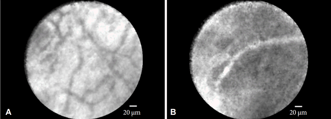
Fig. 1. Probed-based confocal laser endomicroscopy findings in the healthy bile duct. (A) Reticular network of thin dark branching bands (<20 µm) with light gray background. (B) Vessels <20 µm.
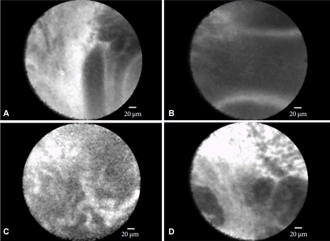
Fig. 2. The Miami classification for malignant pancreaticobiliary strictures. (A) Epithelial structures. (B) Thick dark bands (>40 µm). (C) Thick white bands (>20 µm). (D) Dark clumps.
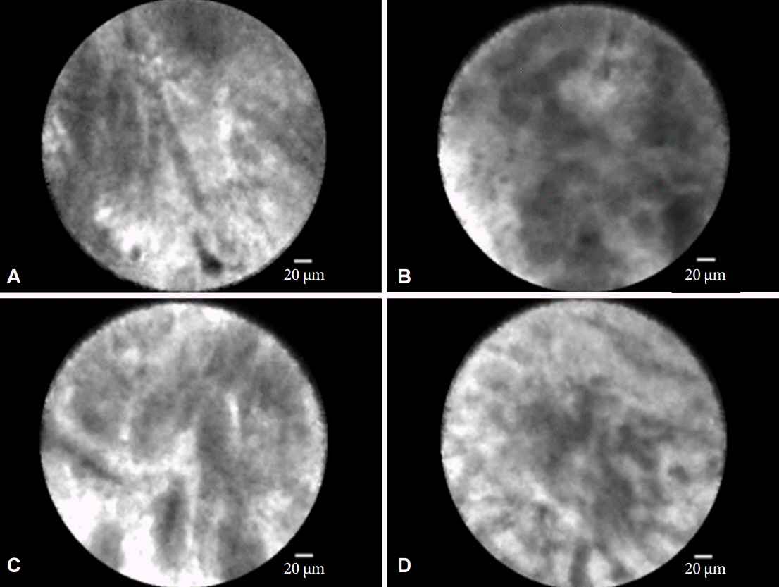
Fig. 3. The Paris classification for inflammatory strictures. (A) Vascular congestion. (B) Dark granular pattern with scales. (C) Increased interglandular space. (D) Thickened reticular structures.

Fig. 4. Needle-based confocal laser endomicroscopy findings with 100% specificity. (A) Villous structures in mucinous pancreatic cystic lesions. (B) Superficial vascular network in serous cystadenomas.
Reference
-
1. Nwaneshiudu A, Kuschal C, Sakamoto FH, Anderson RR, Schwarzenberger K, Young RC. Introduction to confocal microscopy. J Invest Dermatol. 2012; 132:e3.
Article2. Navaneethan U, Njei B, Lourdusamy V, Konjeti R, Vargo JJ, Parsi MA. Comparative effectiveness of biliary brush cytology and intraductal biopsy for detection of malignant biliary strictures: a systematic review and meta-analysis. Gastrointest Endosc. 2015; 81:168–176.
Article3. Slivka A, Gan I, Jamidar P, et al. Validation of the diagnostic accuracy of probe-based confocal laser endomicroscopy for the characterization of indeterminate biliary strictures: results of a prospective multicenter international study. Gastrointest Endosc. 2015; 81:282–290.
Article4. Meining A, Shah RJ, Slivka A, et al. Classification of probe-based confocal laser endomicroscopy findings in pancreaticobiliary strictures. Endoscopy. 2012; 44:251–257.
Article5. Caillol F, Filoche B, Gaidhane M, Kahaleh M. Refined probe-based confocal laser endomicroscopy classification for biliary strictures: the Paris Classification. Dig Dis Sci. 2013; 58:1784–1789.
Article6. Fugazza A, Gaiani F, Carra MC, et al. Confocal laser endomicroscopy in gastrointestinal and pancreatobiliary diseases: a systematic review and meta-analysis. Biomed Res Int. 2016; 2016:4638683.
Article7. Afify AM, al-Khafaji BM, Kim B, Scheiman JM. Endoscopic ultrasound-guided fine needle aspiration of the pancreas. Diagnostic utility and accuracy. Acta Cytol. 2003; 47:341–348.8. Eloubeidi MA, Jhala D, Chhieng DC, et al. Yield of endoscopic ultrasound-guided fine-needle aspiration biopsy in patients with suspected pancreatic carcinoma. Cancer. 2003; 99:285–292.
Article9. Konda VJ, Meining A, Jamil LH, et al. A pilot study of in vivo identification of pancreatic cystic neoplasms with needle-based confocal laser endomicroscopy under endosonographic guidance. Endoscopy. 2013; 45:1006–1013.
Article10. Napoléon B, Lemaistre AI, Pujol B, et al. A novel approach to the diagnosis of pancreatic serous cystadenoma: needle-based confocal laser endomicroscopy. Endoscopy. 2015; 47:26–32.
Article11. Karia K, Waxman I, Konda VJ, et al. Needle-based confocal endomicroscopy for pancreatic cysts: the current agreement in interpretation. Gastrointest Endosc. 2016; 83:924–927.
Article12. Kwan AS, Barry C, McAllister IL, Constable I. Fluorescein angiography and adverse drug reactions revisited: the Lions Eye experience. Clin Experiment Ophthalmol. 2006; 34:33–38.
Article13. Karia K, Saul A, Tyberg A, Gaidhane M, Kahaleh M. Cholangioscopy and biliary confocal laser endoscopy. In : Konda VJ, Waxman I, editors. Endoscopic Imaging Techniques and Tools. New York: Springer;2016.14. Cellvizio . Downstream revenue generated by probe-based confocal laser endomicroscopy (pCLE) in undetermined pancreaticobiliary lesions [Internet]. Paris: Mauna Kea Technologies;c2015 [cited 2016 Aug 9]. Available from: http://www.cellvizio.net/sites/default/files/issu_presentation/ICCU2014%20-%20Sharaiha.pdf.15. Karia K, Gaidhane M, Kahaleh M, Sharaiha RZ. Probe-based confocal laser endomicroscopy for the evaluation of indeterminate biliary strictures: is it cost effective? Gastrointest Endosc. 2016; 83(5 Suppl):AB191.
- Full Text Links
-
- Actions
-
Cited
- CITED
-
- Close
- Share
-
- Similar articles
-
- High-Resolution Probe-Based Confocal Laser Endomicroscopy for Diagnosing Biliary Diseases
- Usefulness of Probe-Based Confocal Laser Endomicroscopy for Esophageal Squamous Cell Neoplasm
- Confocal Laser Endomicroscopy and Molecular Imaging in Barrett Esophagus and Stomach
- Flexible Spectral Imaging Color Enhancement and Probe-based Confocal Laser Endomicroscopy in Minimal Change Esophageal Reflux Disease
- Probe-based confocal laser endomicroscopy in the differential diagnosis of inflammatory bowel diseases: a case series
