Yonsei Med J.
2015 Jan;56(1):182-188. 10.3349/ymj.2015.56.1.182.
Overcoming the Limitations of Fine Needle Aspiration Biopsy: Detection of Lateral Neck Node Metastasis in Papillary Thyroid Carcinoma
- Affiliations
-
- 1Department of Surgery, CHA Bundang Medical Center, CHA University, Seongnam, Korea.
- 2Thyroid Cancer Center, Department of Surgery, Gangnam Severance Hospital, Yonsei University College of Medicine, Seoul, Korea. surghsc@yuhs.ac
- KMID: 2352805
- DOI: http://doi.org/10.3349/ymj.2015.56.1.182
Abstract
- PURPOSE
Ultrasound (US) and US-guided fine needle aspiration biopsies (FNAB) are considered the modalities of choice for assessing lymph nodes suspected of containing metastases, but the sensitivity of FNAB varies and is specific to the operator. We analyzed the risk of FNAB providing false negative results of lateral neck node metastasis, and evaluated diagnostic accuracy of FNAB, in patients with papillary thyroid cancer.
MATERIALS AND METHODS
FNAB was performed in 242 patients suspected of having lateral neck node metastasis on preoperative imaging. Thyroglobulin in the fine-needle aspirate washout (FNA wash-out Tg) and computed tomography enhancement (Hounsfield units) were measured. Patients with negative results on FNAB were examined by intraoperative frozen section. The false negative and true negative groups were compared.
RESULTS
Of the 242 patients, 130 were confirmed as having lateral neck node metastases. In 74 patients, the metastasis was identified by FNAB. False positive results were observed in 2 patients (0.8%) and false negatives in 58 (44.6%). Risk analysis showed that patient age <45 years (p=0.006), tumor size >1 cm (p=0.008) and elevated FNA wash-out Tg (p=0.004) were significantly associated with false negative results on FNAB. The accuracy of FNAB increased significantly when combined with FNA wash-out Tg (p=0.003).
CONCLUSION
To reduce the false negative rate of FNAB, patient age (<45 years), tumor size (>1 cm) and FNA wash-out Tg (>34.8 ng/mL) should be considered in preoperative planning. Accuracy may be improved by combining the results of FNAB and FNA wash-out Tg.
Keyword
MeSH Terms
-
Adolescent
Adult
Aged
Biopsy, Fine-Needle
Carcinoma/*diagnosis/*pathology/radiography/surgery
False Negative Reactions
Female
Humans
Lymph Nodes/*pathology/radiography
Lymphatic Metastasis/*pathology/radiography
Male
Middle Aged
Multivariate Analysis
Preoperative Care
Risk Factors
Sensitivity and Specificity
Thyroglobulin/metabolism
Thyroid Gland/*pathology
Thyroid Neoplasms/*diagnosis/*pathology/radiography/surgery
Tomography, X-Ray Computed
Young Adult
Thyroglobulin
Figure
-
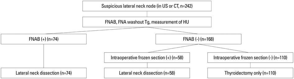
Fig. 1 Diagram of the algorithm for evaluation and management of lateral neck node. FNAB, fine needle aspiration biopsy; US, ultrasound; FNA, fine needle aspiration; Tg, thyroglobulin; HU, Hounsfield unit.
Reference
-
1. Kim E, Park JS, Son KR, Kim JH, Jeon SJ, Na DG. Preoperative diagnosis of cervical metastatic lymph nodes in papillary thyroid carcinoma: comparison of ultrasound, computed tomography, and combined ultrasound with computed tomography. Thyroid. 2008; 18:411–418.
Article2. Mazzaferri EL, Kloos RT. Clinical review 128: Current approaches to primary therapy for papillary and follicular thyroid cancer. J Clin Endocrinol Metab. 2001; 86:1447–1463.
Article3. Ito Y, Miyauchi A. Lateral lymph node dissection guided by preoperative and intraoperative findings in differentiated thyroid carcinoma. World J Surg. 2008; 32:729–739.
Article4. Lim CY, Sohn EJ, Lee J, Yun JS, Nam KH, Chang HS, et al. The significant predicting factors influencing lateral neck node metastasis in papillary thyroid carcinoma. J Korean Surg Soc. 2006; 71:326–330.5. Ito Y, Tomoda C, Uruno T, Takamura Y, Miya A, Kobayashi K, et al. Ultrasonographically and anatomopathologically detectable node metastases in the lateral compartment as indicators of worse relapse-free survival in patients with papillary thyroid carcinoma. World J Surg. 2005; 29:917–920.
Article6. Mazzaferri EL, Young RL. Papillary thyroid carcinoma: a 10 year follow-up report of the impact of therapy in 576 patients. Am J Med. 1981; 70:511–518.
Article7. Shaha AR, Shah JP, Loree TR. Risk group stratification and prognostic factors in papillary carcinoma of thyroid. Ann Surg Oncol. 1996; 3:534–538.
Article8. Steinmüller T, Klupp J, Rayes N, Ulrich F, Jonas S, Gräf KJ, et al. Prognostic factors in patients with differentiated thyroid carcinoma. Eur J Surg. 2000; 166:29–33.
Article9. Cooper DS, Doherty GM, Haugen BR, Kloos RT, Lee SL, Mandel SJ, et al. Management guidelines for patients with thyroid nodules and differentiated thyroid cancer. Thyroid. 2006; 16:109–142.
Article10. Choi JS, Kim J, Kwak JY, Kim MJ, Chang HS, Kim EK. Preoperative staging of papillary thyroid carcinoma: comparison of ultrasound imaging and CT. AJR Am J Roentgenol. 2009; 193:871–878.
Article11. Frasoldati A, Valcavi R. Challenges in neck ultrasonography: lymphadenopathy and parathyroid glands. Endocr Pract. 2004; 10:261–268.
Article12. Frasoldati A, Toschi E, Zini M, Flora M, Caroggio A, Dotti C, et al. Role of thyroglobulin measurement in fine-needle aspiration biopsies of cervical lymph nodes in patients with differentiated thyroid cancer. Thyroid. 1999; 9:105–111.
Article13. Kim MJ, Kim EK, Kim BM, Kwak JY, Lee EJ, Park CS, et al. Thyroglobulin measurement in fine-needle aspirate washouts: the criteria for neck node dissection for patients with thyroid cancer. Clin Endocrinol (Oxf). 2009; 70:145–151.
Article14. Pacini F, Fugazzola L, Lippi F, Ceccarelli C, Centoni R, Miccoli P, et al. Detection of thyroglobulin in fine needle aspirates of nonthyroidal neck masses: a clue to the diagnosis of metastatic differentiated thyroid cancer. J Clin Endocrinol Metab. 1992; 74:1401–1404.
Article15. Sohn YM, Kwak JY, Kim EK, Moon HJ, Kim SJ, Kim MJ. Diagnostic approach for evaluation of lymph node metastasis from thyroid cancer using ultrasound and fine-needle aspiration biopsy. AJR Am J Roentgenol. 2010; 194:38–43.
Article16. Jeong HS, Baek CH, Son YI, Choi JY, Kim HJ, Ko YH, et al. Integrated 18F-FDG PET/CT for the initial evaluation of cervical node level of patients with papillary thyroid carcinoma: comparison with ultrasound and contrast-enhanced CT. Clin Endocrinol (Oxf). 2006; 65:402–407.
Article17. Jeong JJ, Lee YS, Lee SC, Kang SW, Chung WY, Chang HS, et al. A scoring system for prediction of lateral neck node metastasis from papillary thyroid cancer. J Korean Med Sci. 2011; 26:996–1000.
Article18. Mirallié E, Sagan C, Hamy A, Paineau J, Kahn X, Le Néel JC, et al. Predictive factors for node involvement in papillary thyroid carcinoma. Univariate and multivariate analyses. Eur J Cancer. 1999; 35:420–423.
Article19. Yoon JH, Kim JY, Moon HJ, Youk JH, Son EJ, Kim EK, et al. Contribution of computed tomography to ultrasound in predicting lateral lymph node metastasis in patients with papillary thyroid carcinoma. Ann Surg Oncol. 2011; 18:1734–1741.
Article20. American Thyroid Association (ATA) Guidelines Taskforce on Thyroid Nodules and Differentiated Thyroid Cancer. Cooper DS, Doherty GM, Haugen BR, Kloos RT, Lee SL, et al. Revised American Thyroid Association management guidelines for patients with thyroid nodules and differentiated thyroid cancer. Thyroid. 2009; 19:1167–1214.
Article
- Full Text Links
-
- Actions
-
Cited
- CITED
-
- Close
- Share
-
- Similar articles
-
- Oxyphilic Papillary Carcinoma of the Thyroid in Fine Needle Aspiration
- Concurrent Papillary and Medullary Carcinoma of the Thyroid Gland
- Cutaneous Implantation Metastasis of Papillary Thyroid Carcinoma Following Fine Needle Aspiration Biopsy
- Managing Thyroid Microcarcinomas
- Fine needle aspiration cytology of mixed squamous cell carcinoma and papillary carcinoma in thyroid: a case report
