Demineralized dentin matrix combined with recombinant human bone morphogenetic protein-2 in rabbit calvarial defects
- Affiliations
-
- 1R&D Institute, Korea Tooth Bank, Seoul, Korea.
- 2Department of Medicine, Korea University Graduate School, Seoul, Korea.
- 3Department of Oral and Maxillofacial Surgery, Section of Dentistry, Seoul National University Bundang Hospital, Seongnam, Korea.
- 4Department of Oral and Maxillofacial Surgery, College of Dentistry, Dankook University, Cheonan, Korea.
- 5Department of Dentistry, Korea University Anam Hospital, Seoul, Korea. koprosth@unitel.co.kr
- 6Department of Dentistry, Korea University Ansan Hospital, Ansan, Korea. omfs1109@korea.ac.kr
- KMID: 2351731
- DOI: http://doi.org/10.5125/jkaoms.2016.42.2.90
Abstract
OBJECTIVES
The aim of this study was to compare the osteogenic effects of demineralized dentin matrix (DDM) combined with recombinant human bone morphogenetic protein-2 (rhBMP-2) in rabbit calvarial defects with DDM and anorganic bovine bone (ABB) combined with rhBMP-2.
MATERIALS AND METHODS
Four round defects with 8-mm diameters were created in each rabbit calvaria. Each defect was treated with one of the following: 1) DDM, 2) ABB/rhBMP-2, or 3) DDM/rhBMP-2. The rhBMP-2 was combined with DDM and ABB according to a stepwise dry and dip lyophilizing protocol. Histological and microcomputed tomography (µCT) analyses were performed to measure the amount of bone formation and bone volume after 2- and 8-week healing intervals.
RESULTS
Upon histological observation at two weeks, the DDM and ABB/rhBMP-2 groups showed osteoconductive bone formation, while the DDM/rhBMP-2 group showed osteoconductive and osteoinductive bone formation. New bone formation was higher in DDM/rhBMP-2, DDM and ABB decreasing order. The amounts of bone formation were very similar at two weeks; however, at eight weeks, the DDM/rhBMP-2 group showed a two-fold greater amount of bone formation compared to the DDM and ABB/rhBMP-2 groups. The µCT analysis showed markedly increased bone volume in the DDM/rhBMP-2 group at eight weeks compared with that of the DDM group. Notably, there was a slight decrease in bone volume in the ABB/rhBMP-2 group at eight weeks. There were no significant differences among the DDM, ABB/rhBMP-2, and DDM/rhBMP-2 groups at two or eight weeks.
CONCLUSION
Within the limitations of this study, DDM appears to be a suitable carrier for rhBMP-2 in orthotopic sites.
Keyword
Figure
-

Fig. 1 A. Microcomputed tomography image of an en bloc rabbit calvarial defect. Top left: DDM, top right: DDM, bottom left: ABB/rhBMP-2, bottom right: DDM/rhBMP-2. B. Histologic specimen of DDM at two weeks (H&E staining, ×100). Note the defect border, where DDM particles were grafted. (DDM: demineralized dentin matrix, ABB: anorganic bovine bone, rhBMP-2: recombinant human bone morphogenetic protein-2)

Fig. 2 A. Microcomputed tomography image of calvarial defect, cross section at two weeks after demineralized dentin matrix (DDM) implantation. B. Three-dimensional reconstruction of grafted area as volume of interest (8×1 mm) in the form of a disk from the red line (in Fig. 2. A). C. Quantification of new bone volume at two weeks after DDM implantation using NRecon reconstruction software (NRecon v.1.4.4; SkyScan).

Fig. 3 A. DDM at two weeks (H&E staining, ×100). New bone was formed and migrated from the bone defect margin that has abundant osteocytes and blood vessels. All of the fibroblasts around the scaffold and new osteoids were stimulated and transformed phenotypically into osteoblast-like cells. B. ABB/rhBMP-2 at two weeks (H&E staining, ×100). New bone was formed and migrated from the bone defect margin to the surface of the ABB scaffold. The cells around the ABB were stabilized, and spaces were filled with dense fibrous connective tissues. C. DDM/rhBMP-2 at two weeks (H&E staining, ×200). Osteoinductive deposits of osteoids are evident, as is the phenotypic transformation of osteoblast-like cells on the surfaces of the osteoids. There was abundant vascular proliferation as evidence of the remodeling capacity in the newly deposited osteoids on the surface of DDM/rhBMP-2. (DDM: demineralized dentin matrix, ABB: anorganic bovine bone, rhBMP-2: recombinant human bone morphogenetic protein-2)
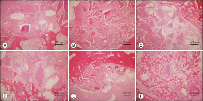
Fig. 4 Histomorphometric measurement of new bone formation at two and eight weeks (H&E staining, ×100). A. DDM at two weeks. B. ABB/rhBMP-2 at two weeks. C. DDM/rhBMP-2 at two weeks. D. DDM at eight weeks. E. ABB/rhBMP-2 at eight weeks. F. DDM/rhBMP-2 at eight weeks. The implantation of scaffolds loaded with rhBMP-2 (DDM and ABB) showed significantly increased new bone formation during the period from two to eight weeks. There were no statistically significant differences at P<0.05. (DDM: demineralized dentin matrix, ABB: anorganic bovine bone, rhBMP-2: recombinant human bone morphogenetic protein-2)
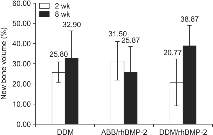
Fig. 5 New bone volume measured by microcomputed tomography. New bone volume increased in DDM (27.5%) and DDM/rhBMP-2 (87.14%) from two to eight weeks compared with the decreased new bone volume in ABB/rhBMP-2 (–17.9%). Values are presented as mean±standard deviation. No statistically significant differences were found among the groups. (DDM: demineralized dentin matrix, ABB: anorganic bovine bone, rhBMP-2: recombinant human bone morphogenetic protein-2)
Reference
-
1. Govender S, Csimma C, Genant HK, Valentin-Opran A, Amit Y, Arbel R, et al. Recombinant human bone morphogenetic protein-2 for treatment of open tibial fractures: a prospective, controlled, randomized study of four hundred and fifty patients. J Bone Joint Surg Am. 2002; 84:2123–2134. PMID: 12473698.2. Burkus JK, Heim SE, Gornet MF, Zdeblick TA. Is INFUSE bone graft superior to autograft bone? An integrated analysis of clinical trials using the LT-Cage lumbar tapered fusion device. J Spinal Disord Tech. 2003; 16:113–122. PMID: 12679664.
Article3. Ripamonti U, Reddi AH. Periodontal regeneration: potential role of bone morphogenetic proteins. J Periodontal Res. 1994; 29:225–235. PMID: 7932015.
Article4. Asahina I. Bone morphogenetic proteins: their history and characteristics. J Hard Tissue Biol. 2014; 23:283–286.
Article5. Gauthier O, Bouler JM, Aguado E, Pilet P, Daculsi G, Macroporous . Biphasic calcium phosphate ceramics: influence of macropore diameter and macroporosity percentage on bone ingrowth. Biomaterials. 1998; 19:133–139. PMID: 9678860.6. Kim CS, Kim JI, Kim J, Choi SH, Chai JK, Kim CK, et al. Ectopic bone formation associated with recombinant human bone morphogenetic proteins-2 using absorbable collagen sponge and beta tricalcium phosphate as carriers. Biomaterials. 2005; 26:2501–2507. PMID: 15585252.
Article7. Hyun SJ, Han DK, Choi SH, Chai JK, Cho KS, Kim CK, et al. Effect of recombinant human bone morphogenetic protein-2, -4, and -7 on bone formation in rat calvarial defects. J Periodontol. 2005; 76:1667–1674. PMID: 16253088.
Article8. Triplett RG, Nevins M, Marx RE, Spagnoli DB, Oates TW, Moy PK, et al. Pivotal randomized, parallel evaluation of recombinant human bone morphogenetic protein-2/absorbable collagen sponge and autogenous bone graft for maxillary sinus floor augmentation. J Oral Maxillofac Surg. 2009; 67:1947–1960. PMID: 19686934.
Article9. Ike M, Urist MR. Recycled dentin root matrix for a carrier of recombinant human bone morphogenetic protein. J Oral Implantol. 1998; 24:124–132. PMID: 9893518.
Article10. Murata M. Bone engineering using human demineralixed dentin matrix and recombinant human BMP-2. J Hard Tissue Biol. 2005; 14:80–81.11. Kim YK, Um IW, An HJ, Kim KW, Hong KS, Murata M. Effects of demineralized dentin matrix used as an rhBMP-2 carrier for bone regeneration. J Hard Tissue Biol. 2014; 23:415–422.
Article12. Kim YK, Kwon KH, Lee ES, Kim CH, Kim MY, Um IW. Experimental study on human demineralized dentin matrix as rhBMP-2 carrier in vivo. J Dent App. 2015; 2:269–273.13. Jung JH, Yun JH, Um YJ, Jung UW, Kim CS, Choi SH, et al. Bone formation of Escherichia coli expressed rhBMP-2 on absorbable collagen block in rat calvarial defects. Oral Surg Oral Med Oral Pathol Oral Radiol Endod. 2011; 111:298–305. PMID: 20875759.
Article14. Geiger M, Li RH, Friess W. Collagen sponges for bone regeneration with rhBMP-2. Adv Drug Deliv Rev. 2003; 55:1613–1629. PMID: 14623404.
Article15. Fiorellini JP, Howell TH, Cochran D, Malmquist J, Lilly LC, Spagnoli D, et al. Randomized study evaluating recombinant human bone morphogenetic protein-2 for extraction socket augmentation. J Periodontol. 2005; 76:605–613. PMID: 15857102.
Article16. McKay WF, Peckham SM, Marotta JS. The science of rhBMP-2. St. Louis: Quality Medical Publishing;2006.17. Schützenberger S, Schultz A, Hausner T, Hopf R, Zanoni G, Morton T, et al. The optimal carrier for BMP-2: a comparison of collagen versus fibrin matrix. Arch Orthop Trauma Surg. 2012; 132:1363–1370. PMID: 22660797.
Article18. Jung SY, KO YJ, Jang HS, Kang SW, Park JH. The effect of carrier for BMP-2 delivery on histological aspects of tissue-engineered bone. Tissue Eng Regen Med. 2013; 10:341–346.
Article19. Schmitt C, Lutz R, Doering H, Lell M, Ratky J, Schlegel KA. Bio-Oss block combined with BMP-2 and VEGF for the regeneration of bony defects and vertical augmentation. Clin Oral Implants Res. 2013; 24:450–460. PMID: 22092937.20. Kim YK, Um IW, Murata M. Tooth bank system for bone regeneration: safety report. J Hard Tissue Biol. 2014; 23:371–376.21. Kim YK, Kim SG, Byeon JH, Lee HJ, UM IU, Lim SC, et al. Development of a novel bone grafting material using autogenous teeth. Oral Surg Oral Med Oral Pathol Oral Radiol Endod. 2010; 109:496–503. PMID: 20060336.
Article22. Murata M, Kawai T, Kawakami T, Akazawa T, Tazaki J, Ito K, et al. Human acid-insoluble dentin with BMP-2 accelerates bone induction in subcutaneous and intramuscular tissues. J Ceram Soc Jpn. 2010; 118:438–441.
Article
- Full Text Links
-
- Actions
-
Cited
- CITED
-
- Close
- Share
-
- Similar articles
-
- Bone regeneration of demineralized dentin matrix with platelet-rich fibrin and recombinant human bone morphogenetic protein-2 on the bone defects in rabbit calvaria
- Bone Induction by Demineralized Dentin Matrix in Nude Mouse Muscles
- Postulated release profile of recombinant human bone morphogenetic protein-2 (rhBMP-2) from demineralized dentin matrix
- Cranial bone regeneration according to different particle sizes and densities of demineralized dentin matrix in the rabbit model
- Effect of bone wax plus bone morphogenetic protein into rabbit calvarial defects
