Asymptomatic Primary Isolated Pulmonary Vein Stenosis in an Adult: A Case Report
- Affiliations
-
- 1Department of Internal Medicine, College of Medicine, Soonchunhyang University, Cheonan, Korea. khseo@schmc.ac.kr
- 2Department of Diagnostic Radiology, College of Medicine, Soonchunhyang University, Cheonan, Korea.
Abstract
- A 31-year-old man without respiratory symptoms was transferred to our clinic with incidentally detected small nodular densities in both the upper lung zones on chest radiography. Chest computed tomography and pulmonary angiography demonstrated that the entrance of the right inferior pulmonary vein to the left atrium was completely blocked, and the venous return of the right lower lobe was achieved through the right superior pulmonary vein with a tortuous venous collateral complex in the venous phase. With echocardiography, mild pulmonary hypertension was detected. Here, we present an asymptomatic adult with isolated stenosis of the pulmonary vein with chronic compensation by venous collateral circulation in spite of mild pulmonary hypertension.
MeSH Terms
Figure
-
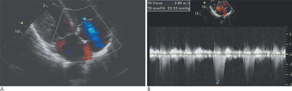
Fig. 1 Echocardiogram shows only turbulent jet flows (arrow) originating from the pulmonary veins and entering the left atrium on apical four chamber view in figure 1A and mild pulmonary hypertension with 38.3 mmHg systolic pulmonary artery pressure (RA pressure 5 mmHg + maximal pressure gradient 33.3 mmHg) in figure 1B.
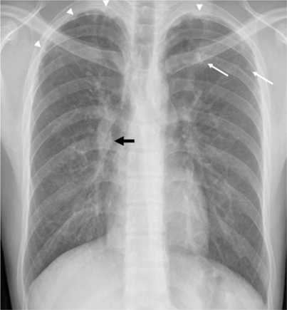
Fig. 2 Chest radiograph shows small nodular densities (arrows) with pleural thickening (arrow heads) in both upper lung zones and a dilated pulmonary vascular structure in the right hilar region (black arrow).
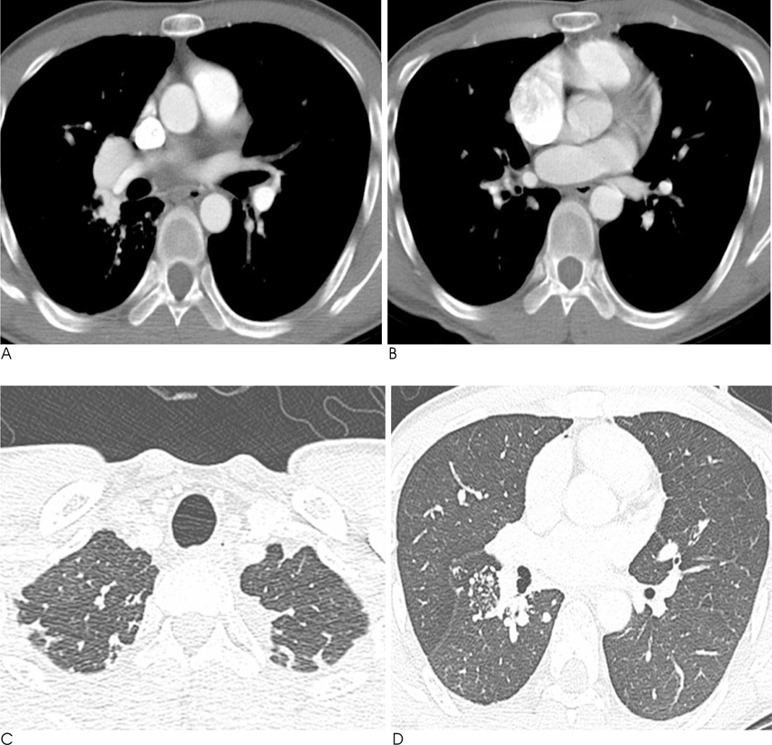
Fig. 3 Contrast-enhanced CT scan shows markedly a dilated right superior pulmonary vein (A) without connection to the left atrium (B), and multiple nodules suggesting collateral vascular structures in the pleural area of both upper lung zones (C) and in the superior segment of right lower lobe (D).

Fig. 4 Volume rendering image shows that the right inferior pulmonary vein drains to the superior pulmonary vein an through intrapulmonary venous connection (arrows).

Fig. 5 Selective angiogram of right lower lobar pulmonary artery shows that the entrance of the right inferior pulmonary vein to the left atrium was completely blocked (arrow), and the venous return of right lower lobe was achieved through the right superior pulmonary vein with tortuous venous collateral complex around the right hilar region in venous phase.
Reference
-
1. Edward J. Congenital stenosis of the pulmonary veins: pathologic and developmental considerations. Lab Invest. 1960; 9:46–66.2. Omasa M, Hasegawa S, Bando T, Okano Y, Otani H, Nakshima Y, et al. A case of congenital pulmonary vein stenosis in an adult. Respiration. 2004; 71:92–94.3. Tan CW, Munfakh N, Helmcke F, Abourahma A, Caspi J, Glancy DL. Congenital bilateral pulmonary venous stenosis in an adult: diagnosis by echo-Doppler. Catheter Cardiovasc Interv. 2000; 49:328–330.4. Saad EB, Marrouche NF, Saad CP, Ha E, Bash D, White RD, et al. Pulmonary vein stenosis after catheter ablation of atrial fibrillation: emergence of a new clinical syndrome. Ann Intern Med. 2003; 138:634–638.5. Van Son JA, Danielson GK, Puga FJ, Edwards WD, Driscoll DJ. Repair of congenital and acquired pulmonary vein stenosis. Ann Thorac Surg. 1995; 60:144–150.6. Latson LA, Prieto LR. Congenital and acquired pulmonary vein stenosis. Circulation. 2007; 115:103–108.7. Breinholt JP, Hawkins JA, Minich L, Tani LY, Orsmond GS, Ritter S, et al. Pulmonary vein stenosis with normal connection: associated cardiac abnormalities and variable outcome. Ann Thorac Surg. 1999; 68:164–168.8. Sun CC, Doyle T, Ringer RE. Pulmonary vein stenosis. Hum Pathol. 1995; 26:880–886.9. Belcourt CL, Roy DL, Nanton MA, Finley JP, Gillis DA, Krause VW, et al. Stenosis of individual pulmonary veins: radiologic findings. Radiology. 1986; 161:109–112.10. Saida Y, Eguchi N, Mori K, Tanaka YO, Ishikawa S, Itai Y. Isolated pulmonary vein stenosis associated with full intrapulmonary compensation. AJR Am J Roentgenol. 1999; 173:961–962.
- Full Text Links
-
- Actions
-
Cited
- CITED
-
- Close
- Share
-
- Similar articles
-
- Cystic Lung Changes in a Thin Section CT in an Asymptomatic Young Adult with Unilateral Pulmonary Vein Atresia: A Case Report
- Unilateral Pulmonary Vein Atresia: A Case Report
- Pulsed Wave and Color Doppler Echocardiography and Cardiac Catheterization Findings in Bilateral Pulmonary Vein Stenosis
- Pulmonary Vein Varix: A Case Report
- Percutaneous Pulmonary Vein Angioplasty for the Pulmonary Vein Stenosis After Catheter Ablation of Atrial Fibrillation
