Increased Infiltration of CD8⺠T Cells by Dacarbazine in a Patient with Mucosal Penile Melanoma Refractory to Nivolumab
- Affiliations
-
- 1Department of Dermatology, Graduate School of Medical and Dental Sciences, Tokyo Medical and Dental University, Tokyo, Japan. tnamderm@tmd.ac.jp
- 2Department of Plastic Surgery, Graduate School of Medical and Dental Sciences, Tokyo Medical and Dental University, Tokyo, Japan.
- 3Department of Pathology, Graduate School of Medical and Dental Sciences, Tokyo Medical and Dental University, Tokyo, Japan.
- KMID: 2344820
- DOI: http://doi.org/10.5021/ad.2016.28.4.486
Abstract
- Primary penile melanomas are rare tumors that represent less than 0.1% of all melanomas. We report a case of a 60-year-old Japanese male with a mucosal penile melanoma and describe an increased CD8⺠T cell infiltration in brain after dacarbazine (DTIC) administration. After partial penectomy and left inguinal lymphadenectomy, he developed multiple lung, bone, spleen, brain and skin metastases. He was treated with interferon-β, DTIC and nivolumab. However, the metastases were not reduced in size. Immunohistochemistry showed an increase of CD8⺠T cell infiltration and programmed death-ligand 1 (PD-L1) expression after the administration of DTIC, but the expression of programmed cell death protein 1 (PD-1) was negative. We speculate that DTIC exerted immunostimulatory effects, but nivolumab was ineffective due to the negative expression of PD-1 and/or an insufficient infiltration of CD8⺠T cells. Although this is only one case, this case report could be the first step to discuss the development of effective therapies against melanoma to take advantage of the increased CD8⺠T cell infiltration elicited by chemotherapeutic agents. It would be beneficial to pay more attention to the relationship between DTIC and immune checkpoint modulators.
Keyword
MeSH Terms
Figure
-
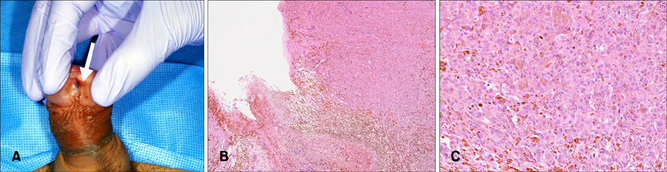
Fig. 1 (A) A pigmented nodule on the urethral orifice (indicated by the white arrow). (B) An atypical melanocytic proliferation arranged in sheets and in nests. (H&E, ×40). (C) Eosinophilic cytoplasm and vesicular pleomorphic nuclei with prominent nucleoli and frequent mitoses (H&E, ×200).
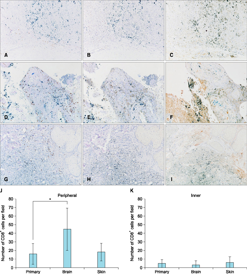
Fig. 2 Immunohistochemistry for CD8 in the primary tumor (A), the brain metastatic lesion (D) and a skin metastatic lesion (G). Immunohistochemistry for programmed cell death protein 1 in the primary tumor (B), the brain metastatic lesion (E) and a skin metastatic lesion (H). Immunohistochemistry for programmed death-ligand 1 in the primary tumor (C), the brain metastatic lesion (F) and a skin metastatic lesion (I) (A~I: ×100). Number of cells staining for CD8 expressed as means±standard deviations measured over 10 high-power fields (×400) in the peripheral (J) and inner (K) layer of the tumor. Counting was performed independently by two observers. *p<0.0001.
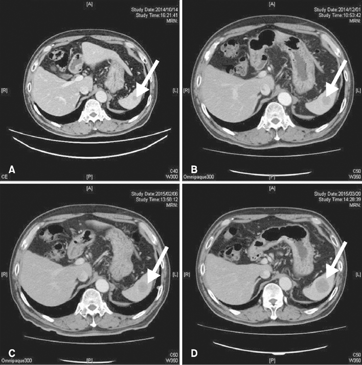
Fig. 3 Contrast-enhanced computed tomography for a splenic metastatic lesion (A) before dacarbazine administration, after dacarbazine administration (B), after the first course of nivolumab administration (C), and after the second course of nivolumab administration (D). Diameters of the splenic metastatic lesion (indicated by white arrows) are 13 mm (A), 20 mm (B), 21 mm (C) and 32 mm (D), respectively. Arrows indicate splenic metastatic lesions.
Reference
-
1. Oliva E, Quinn TR, Amin MB, Eble JN, Epstein JI, Srigley JR, et al. Primary malignant melanoma of the urethra: a clinicopathologic analysis of 15 cases. Am J Surg Pathol. 2000; 24:785–796.2. Ugurel S, Paschen A, Becker JC. Dacarbazine in melanoma: from a chemotherapeutic drug to an immunomodulating agent. J Invest Dermatol. 2013; 133:289–292.
Article3. Müller P, Martin K, Theurich S, von Bergwelt-Baildon M, Zippelius A. Cancer chemotherapy agents target intratumoral dendritic cells to potentiate antitumor immunity. Oncoimmunology. 2014; 3:e954460.
Article4. Hervieu A, Rébé C, Végran F, Chalmin F, Bruchard M, Vabres P, et al. Dacarbazine-mediated upregulation of NKG2D ligands on tumor cells activates NK and CD8 T cells and restrains melanoma growth. J Invest Dermatol. 2013; 133:499–508.
Article5. Hirano F, Kaneko K, Tamura H, Dong H, Wang S, Ichikawa M, et al. Blockade of B7-H1 and PD-1 by monoclonal antibodies potentiates cancer therapeutic immunity. Cancer Res. 2005; 65:1089–1096.6. Ozawa A, Nomiyama T, Nakai N, Hartmann G, Takenaka H, Kishimoto S, et al. Immunohistological analysis of in-transit metastasis in a patient with advanced melanoma treated with combination therapy of cytosine guanine dinucleotide oligodeoxynucleotide, dacarbazine and beta-interferon: a case report. J Dermatol. 2012; 39:1035–1037.
Article7. Nardin A, Wong WC, Tow C, Molina TJ, Tissier F, Audebourg A, et al. Dacarbazine promotes stromal remodeling and lymphocyte infiltration in cutaneous melanoma lesions. J Invest Dermatol. 2011; 131:1896–1905.
Article8. Taube JM, Anders RA, Young GD, Xu H, Sharma R, McMiller TL, et al. Colocalization of inflammatory response with B7-h1 expression in human melanocytic lesions supports an adaptive resistance mechanism of immune escape. Sci Transl Med. 2012; 4:127ra37.
Article
- Full Text Links
-
- Actions
-
Cited
- CITED
-
- Close
- Share
-
- Similar articles
-
- Lichenoid Drug Eruption Developed in Melanoma Patient Treated with Nivolumab
- A Curious Case of Primary Gastric Mucosal Melanoma
- The Major Role of NF-κB in the Depth of Invasion on Acral Melanoma by Decreasing CD8⺠T Cells
- Extrinsic Acquisition of CD80 by Antigen-Specific CD8⺠T Cells Regulates Their Recall Immune Responses to Acute Viral Infection
- Untrastructure of Melanocyte in Penile Melanosis
