Analysis of Protrusio Acetabuli Using a CT-based Diagnostic Method in Korean Patients with Marfan Syndrome: Prevalence and Association with Other Manifestations
- Affiliations
-
- 1Division of Cardiology, Department of Medicine, Heart Vascular Stroke Institute, Samsung Medical Center, Sungkyunkwan University School of Medicine, Seoul, Korea. dukkyung.kim@gmail.com
- 2Department of Critical Care Medicine, Samsung Medical Center, Sungkyunkwan University School of Medicine, Seoul, Korea.
- 3Department of Ophthalmology, Samsung Medical Center, Sungkyunkwan University School of Medicine, Seoul, Korea.
- 4Department of Pediatrics, Samsung Medical Center, Sungkyunkwan University School of Medicine, Seoul, Korea.
- 5Department of Laboratory Medicine and Genetics, Samsung Medical Center, Sungkyunkwan University School of Medicine, Seoul, Korea.
- 6Department of Thoracic and Cardiovascular Surgery, Samsung Medical Center, Sungkyunkwan University School of Medicine, Seoul, Korea.
- 7Department of Radiology, Cardiovascular and Stroke Imaging Center, Samsung Medical Center, Sungkyunkwan University School of Medicine, Seoul, Korea.
- KMID: 2344150
- DOI: http://doi.org/10.3346/jkms.2015.30.9.1260
Abstract
- A new CT-based diagnostic method of protrusio acetabuli (PA) was introduced. However, prevalence of PA by this method and correlation between PA and other manifestations of Marfan syndrome (MFS) is unknown in Korean MFS patients. This study aimed to investigate the prevalence of PA diagnosed by a CT-based method in Korean patients with MFS, the association of PA with other manifestations of MFS, and the contribution of PA to MFS diagnosis. We retrospectively reviewed the records of 146 MFS patients with the presence of a causative FBN1 mutation and 146 age- and sex-matched controls from a single tertiary care center. All MFS patients underwent a complete assessment of criteria based on the revised Ghent nosology. PA was assessed quantitatively using a CT-based circle-wall distance (CWD) method. PA was diagnosed in 77.4% of patients in the MFS group and in 11.0% of the control group. CWD was significantly different between the two groups (1.50 mm vs. -0.64 mm, P<0.001). The presence of PA did not correlate with the presence of ectopia lentis, aortic root diameter, or history of aortic dissection. The presence of PA did not have a significant impact on the final diagnosis of MFS. Even though the presence of PA does not related to the cardinal clinical features of MFS or influence MFS diagnosis, its presence may be helpful for the suspicion of MFS when aortic dissection or aneurysm is found on CT angiography of the aorta because of the high frequency of PA in MFS patients.
MeSH Terms
-
Acetabulum/*abnormalities/radiography
Adolescent
Adult
Aged
Aged, 80 and over
Aortic Aneurysm/*epidemiology/radiography
Comorbidity
Female
Humans
Male
Marfan Syndrome/*epidemiology/*radiography
Middle Aged
Prevalence
Reproducibility of Results
Republic of Korea/epidemiology
Risk Factors
Sensitivity and Specificity
Tomography, X-Ray Computed/*methods/*statistics & numerical data
Young Adult
Figure
-
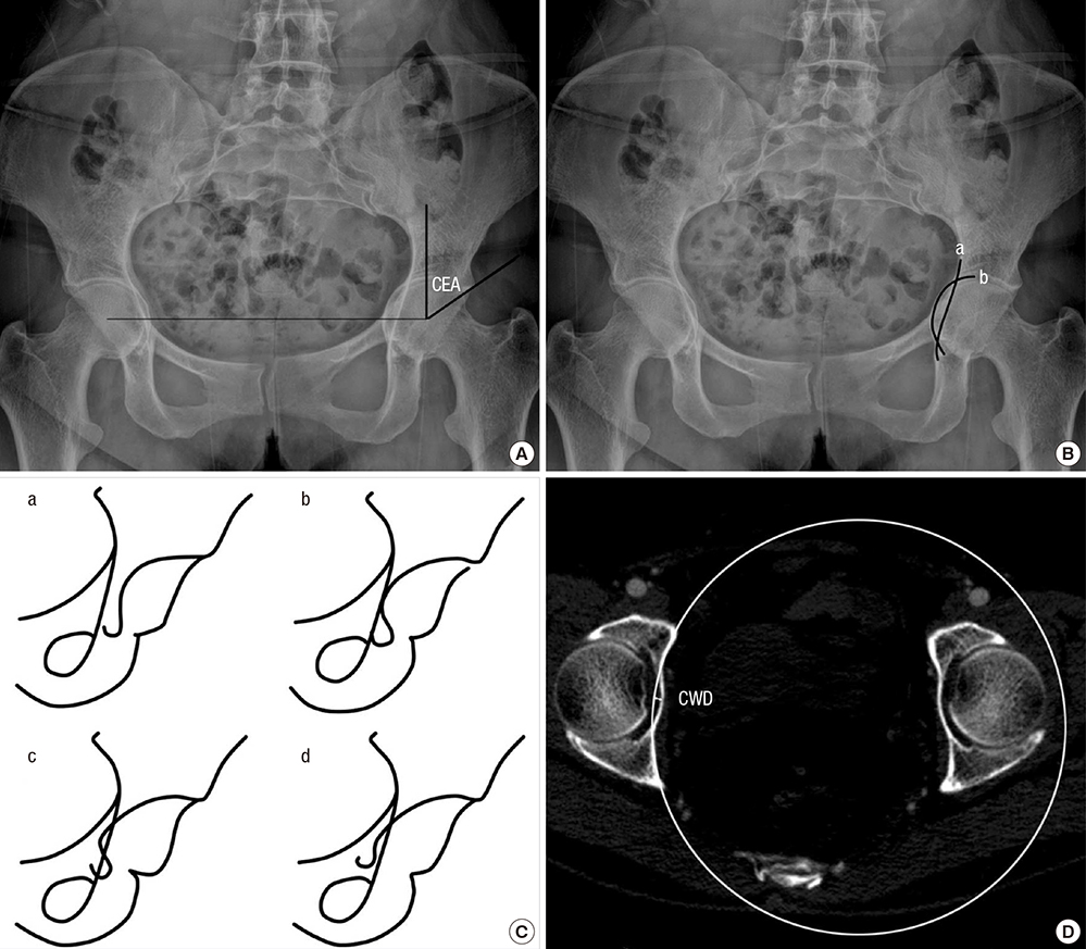
Fig. 1 Diagnostic methods for protrusio acetabuli using plain anteroposterior radiographs of the pelvis and CT images. (A) The center-edge angle (CEA) is formed by a vertical line drawn through the center of the femoral head and a line drawn from the center through the lateral edge of the acetabular roof. A CEA of >50° is considered indicative of protrusio acetabuli. (B) The acetabular-ilioischial distance represents the transverse distance between the ilioischial line (a) and the acetabular line (b). Crossing of the ilioischial line by the acetabular line by > 3 mm medially in men or > 6 mm in women is considered indicative of protrusio acetabuli. (C) Radiographic changes of the teardrop figure in protrusio acetabuli. a, opened; b, closed; c, crossed; d, reversed. (D) CT-based circle-wall distance (CWD) method. A 10 cm radius circle is adapted to the inner acetabular wall of the pelvis. The distance between the line of the circle and the medial most point of the inner pelvic wall of the acetabular fossa was measured. Measurement of CWD is indicated. CT, computed tomography.

Fig. 2 Scattergram of CWD in patients with MFS and normal controls. CWD, circle-wall distance; MFS, Marfan syndrome.
Reference
-
1. Koh KK, Hyon MS, Lim HJ, Kim CH, Oh BH, Park YB, Choi YS, Seo JD, Lee YW. Cardiovascular manifestations of marfan syndrome. Korean Circ J. 1987; 17:777–782.2. Loeys BL, Dietz HC, Braverman AC, Callewaert BL, De Backer J, Devereux RB, Hilhorst-Hofstee Y, Jondeau G, Faivre L, Milewicz DM, et al. The revised Ghent nosology for the Marfan syndrome. J Med Genet. 2010; 47:476–485.3. De Paepe A, Devereux RB, Dietz HC, Hennekam RC, Pyeritz RE. Revised diagnostic criteria for the Marfan syndrome. Am J Med Genet. 1996; 62:417–426.4. McBride MT, Muldoon MP, Santore RF, Trousdale RT, Wenger DR. Protrusio acetabuli: diagnosis and treatment. J Am Acad Orthop Surg. 2001; 9:79–88.5. Steel HH. Protrusio acetabuli: its occurrence in the completely expressed Marfan syndrome and its musculoskeletal component and a procedure to arrest the course of protrusion in the growing pelvis. J Pediatr Orthop. 1996; 16:704–718.6. Do T, Giampietro PF, Burke SW, Davis JG, Raggio C, Schneider R, Boachie-Adjei O, Brill P. The incidence of protrusio acetabuli in Marfan's syndrome and its relationship to bone mineral density. J Pediatr Orthop. 2000; 20:718–721.7. Sohn GH, Jang SY, Moon JR, Yang JH, Sung K, Ki CS, Oh JK, Choe YH, Kim DK. The usefulness of multidetector computed tomographic angiography for the diagnosis of Marfan syndrome by Ghent criteria. Int J Cardiovasc Imaging. 2011; 27:679–688.8. Yang JH, Han H, Jang SY, Moon JR, Sung K, Chung TY, Lee HJ, Ki CS, Kim DK. A comparison of the Ghent and revised Ghent nosologies for the diagnosis of Marfan syndrome in an adult Korean population. Am J Med Genet A. 2012; 158A:989–995.9. Sponseller PD, Jones KB, Ahn NU, Erkula G, Foran JR, Dietz HC 3rd. Protrusio acetabuli in Marfan syndrome: age-related prevalence and associated hip function. J Bone Joint Surg Am. 2006; 88:486–495.10. Armbuster TG, Guerra J Jr, Resnick D, Goergen TG, Feingold ML, Niwayama G, Danzig LA. The adult hip: an anatomic study. Part I: the bony landmarks. Radiology. 1978; 128:1–10.11. Van de Velde S, Fillman R, Yandow S. Protrusio acetabuli in Marfan syndrome. History, diagnosis, and treatment. J Bone Joint Surg Am. 2006; 88:639–646.12. Richards PJ, Pattison JM, Belcher J, Decann RW, Anderson S, Wynn-Jones C. A new tilt on pelvic radiographs: a pilot study. Skeletal Radiol. 2009; 38:113–122.13. Lundby R, Kirkhus E, Rand-Hendriksen S, Hald J, Pripp AH, Smith HJ. CT of the hips in the investigation of protrusio acetabuli in Marfan syndrome. A case control study. Eur Radiol. 2011; 21:1485–1491.14. Sheikhzadeh S, Sondermann C, Rybczynski M, Habermann CR, Brockstaedt L, Keyser B, Kaemmerer H, Mir T, Staebler A, Robinson PN, et al. Comprehensive analysis of dural ectasia in 150 patients with a causative FBN1 mutation. Clin Genet. 2014; 86:238–245.15. Wenger DR, Ditkoff TJ, Herring JA, Mauldin DM. Protrusio acetabuli in Marfan's syndrome. Clin Orthop Relat Res. 1980; 147:134–138.16. Akutsu K, Morisaki H, Takeshita S, Ogino H, Higashi M, Okajima T, Yoshimuta T, Tsutsumi Y, Nonogi H, Morisaki T. Characteristics in phenotypic manifestations of genetically proved Marfan syndrome in a Japanese population. Am J Cardiol. 2009; 103:1146–1148.17. Yoo EH, Woo H, Ki CS, Lee HJ, Kim DK, Kang IS, Park P, Sung K, Lee CS, Chung TY, et al. Clinical and genetic analysis of Korean patients with Marfan syndrome: possible ethnic differences in clinical manifestation. Clin Genet. 2010; 77:177–182.18. Fattori R, Nienaber CA, Descovich B, Ambrosetto P, Reggiani LB, Pepe G, Kaufmann U, Negrini E, von Kodolitsch Y, Gensini GF. Importance of dural ectasia in phenotypic assessment of Marfan's syndrome. Lancet. 1999; 354:910–913.
