Treatment of Acute Central Retinal Artery Occlusion with Ocular Ischemic Syndrome
- Affiliations
-
- 1Department of Ophthalmology, Gachon University Gil Hospital, Incheon, Korea. dylee@gilhospital.com
Abstract
- PURPOSE
We report a case of treatment of acute central retinal artery occlusion (CRAO) with ocular ischemic syndrome (OIS).
CASE SUMMARY
A 72-year-old man presented with acute loss of vision in the right eye on that day. At initial examination, visual acuity tested positive for light sensitivity in the right eye. Fundus examination demonstrated a visible embolus at the central retinal artery overlying the optic disc head and a cherry-red spot in the fovea. Fluorescein angiography revealed that filling of the choroidal circulation was delayed, and the arteriovenous transit time was even further delayed. Carotid angiography showed marked stenosis within the right internal carotid artery. Laboratory tests included blood tests for hypercoagulability evaluation, for which the results were non-specific. To treat acute CRAO with OIS in the right eye, transluminal Nd:YAG laser embolectomy (TYE) was performed twice, and carotid angioplasty with stenting was conducted within the stenosed internal carotid artery. One month after the TYE procedure and carotid stenting, the patient's visual acuity improved to 0.06 and the arteriovenous transit time was within normal limits on fluorescein angiography.
CONCLUSIONS
The visual prognosis in eyes with CRAO plus an associated choroidal circulatory disturbance is extremely poor. However, we experienced and reported a case of CRAO with OIS treated successfully through a prompt TYE procedure and carotid angioplasty with stenting.
Keyword
MeSH Terms
Figure
-
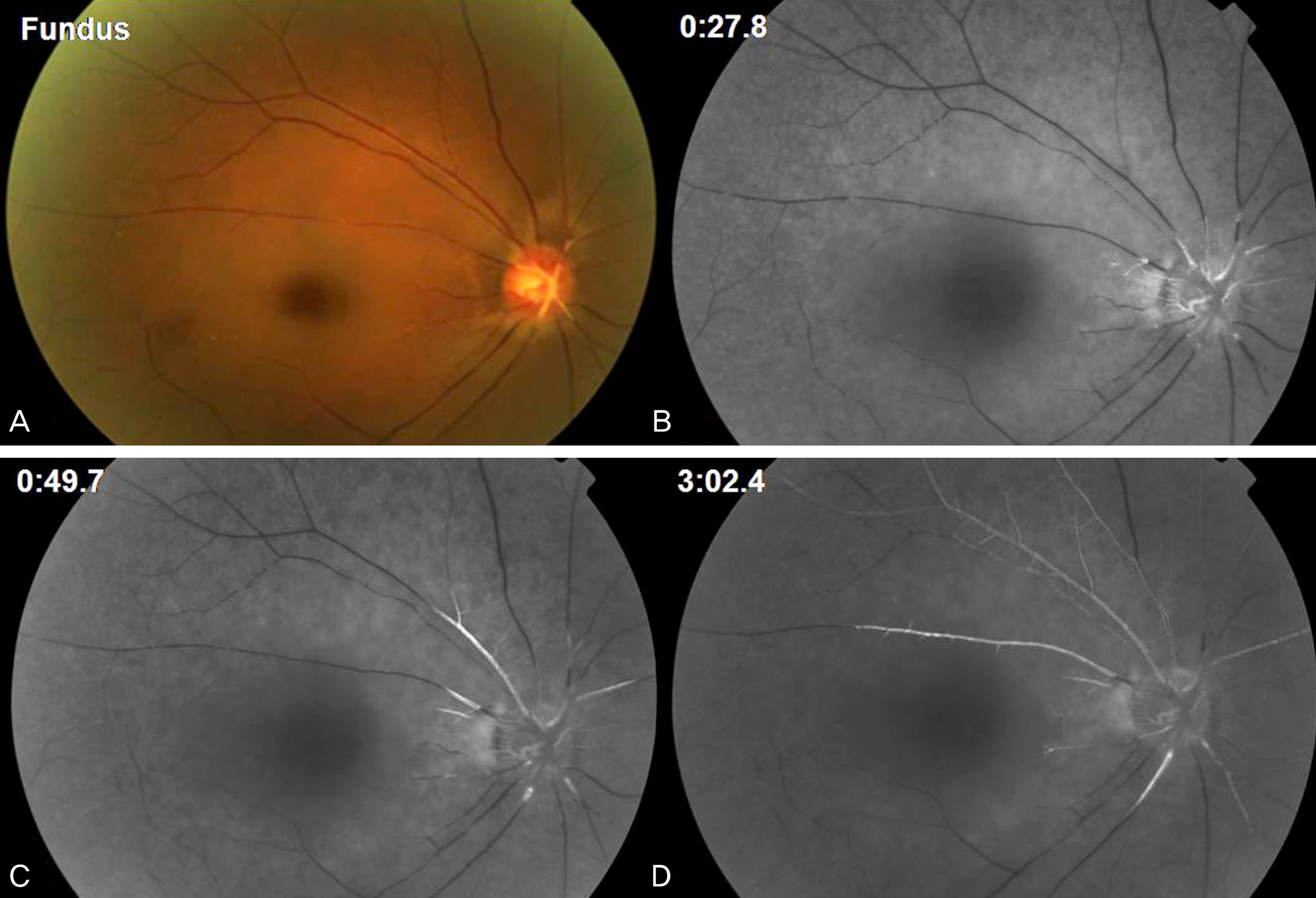
Figure 1. (A) Fundus photograph of the right eye at the first visit. Large emboli were seen at the central retinal artery overlying the optic disc head. Superficial retinal whitening or opacification was noted in the posterior pole with evidence of a cherry-red spot in the fovea. (B, C, D) Fluorescein angiographs of the right eye at the first visit. A filling of the choroidal circulation was delayed and the arteriovenous transit time was even further delayed.

Figure 2. (A) Fundus photograph of the right eye on the third days after first transluminal Nd:YAG laser embolectomy (TYE). Large emboli were not seen at the central retinal artery overlying the optic disc head any more. Vitreous hemorrhages were seen at optic disc area and inferior pole. (B, C, D) Fluorescein angiographs of the right eye on the third days after first TYE. A filling of the choroidal circulation was still delayed. But the arteriovenous transit time was within normal limit.
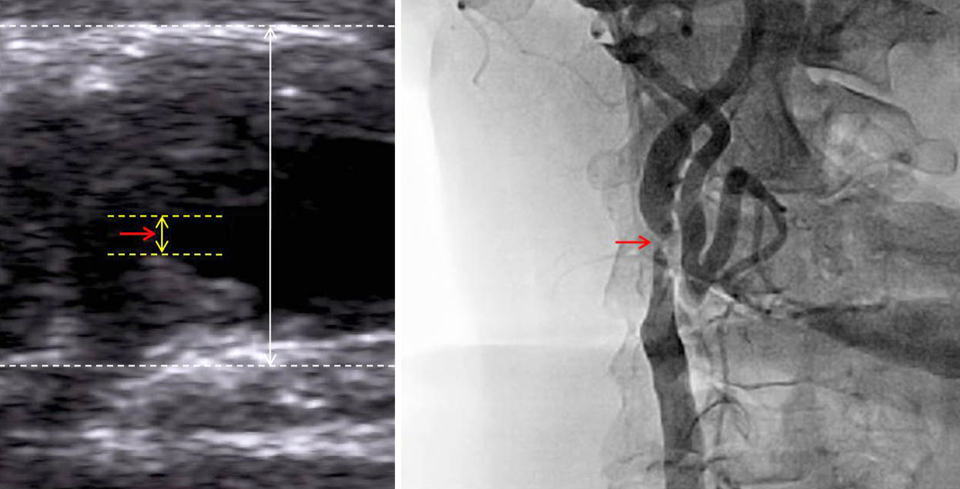
Figure 3. Carotid Doppler sonogram and carotid angiograph of the right eye. A 90% obstruction of the right internal carotid artery was presented (red arrow).
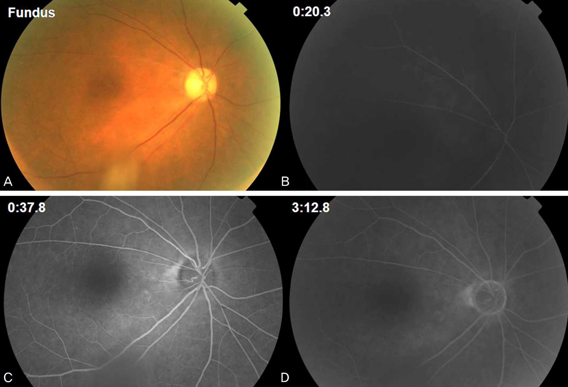
Figure 4. Fundus photograph and fluorescein angiographs of the right eye on the first month after transluminal Nd:YAG laser embolectomy (TYE) procedure and carotid stenting. (A) On fundus photograph, the central retinal artery and retinal arterioles appeared patent and no emboli were seen. (B, C, D) On fluorescein angiographs, the arteriovenous transit time was within normal limit on fluorescein angiography.
Reference
-
References
1. Brown GC, Magargal LE. Central retinal artery obstruction and visual acuity. Ophthalmology. 1982; 89:14–9.
Article2. Biousse V, Calvetti O, Bruce BB, Newman NJ. Thrombolysis for central retinal artery occlusion. J Neuroophthalmol. 2007; 27:215–30.
Article3. Brown GC, Magargal LE, Shields JA, et al. Retinal arterial obstruction in children and young adults. Ophthalmology. 1981; 88:18–25.
Article4. Park JY, Koo NK, Seo WM. Spectral-domain optical coherence tomography findings in acute central retinal artery occlusion. J Korean Ophthalmol Soc. 2012; 53:1099–103.
Article5. Lee SB, Yun YJ, Kim JY. Central retinal artery obstruction in protein S deficiency. J Korean Ophthalmol Soc. 2008; 49:2017–20.
Article6. Lee SJ, Kim SY, Kim SD. Two cases of branch retinal arterial occlusion after carotid artery stenting in the carotid stenosis. Korean Ophthalmol. 2009; 23:53–6.
Article7. Shin JH, Kim DK, Yu SY, Kwak HW. A case of central retinal artery occlusion after intravitreal triamcinolone acetonide injection for diabetic macular edema in non-proliferative diabetic retinopathy. J Korean Ophthalmol Soc. 2006; 47:667–71.8. Opremcak E, Rehmar AJ, Ridenour CD, et al. Restoration of retinal blood flow via translumenal Nd:YAG embolysis/embolectomy (TYL/E) for central and branch retinal artery occlusion. Retina. 2008; 28:226–35.
Article9. Lim JY, Lee JY, Chung HW, et al. Treatment of branch retinal ar-tery occlusion with transluminal Nd:YAG laser embolysis. Korean J Ophthalmol. 2009; 23:315–7.
Article10. Meyer CH, Holz FG. Images in clinical medicine. Blurred vision after cardiac catheterization. N Engl J Med. 2009; 361:2366.11. Feist RM, Emond TL. Translumenal Nd:YAG laser embolysis for central retinal artery occlusion. Retina. 2005; 25:797–9.
Article12. Kearns TP, Hollenhorst RW. Venous-stasis retinopathy of occlusive disease of the carotid artery. Proc Staff Meet Mayo Clin. 1963; 38:304–12.13. Brown GC, Magargal LE. The ocular ischemic syndrome. Clinical, fluorescein angiographic and carotid angiographic features. Int Ophthalmol. 1988; 11:239–51.14. Sivalingam A, Brown GC, Magargal LE. The ocular ischemic syndrome. III. Visual prognosis and the effect of treatment. Int Ophthalmol. 1991; 15:15–20.
Article15. Han YS, Yoo WS, Chung IY, Park JM. Ocular ischemic syndrome successfully treated with carotid angioplasty and stenting. J Korean Ophthalmol Soc. 2010; 51:447–52.
Article16. Wang YL, Zhao L, Li MM. Improved circulation in ocular ischemic syndrome after carotid artery stenting. Chin Med J (Engl). 2011; 124:3598–600.17. Neroev VV, Kiseleva TN, Vlasov SK, et al. Visual outcomes after carotid reconstructive surgery for ocular ischemia. Eye (Lond). 2012; 26:1281–7.
Article
- Full Text Links
-
- Actions
-
Cited
- CITED
-
- Close
- Share
-
- Similar articles
-
- A Case of Ocular Ischemic Syndrome Associated with Multiple Branch Retinal Artery Occlusion and Neovascular Glaucoma
- A Case of Ocular Ischemic Syndrome with Neovascular Glaucoma
- Incomplete Central Retinal Artery Occlusion
- Central Retinal Artery Occlusion after Cervical Spine Surgery in Prone Position: A Case Report
- The Successful Treatment of a Case of Central Retinal Artery Occlusion
