J Korean Ophthalmol Soc.
2014 Jan;55(1):133-137.
A Case of Long Anterior Lens Zonule and Pigment Dispersion Syndrome
- Affiliations
-
- 1Department of Ophthalmology, Jeju National University School of Medicine, Jeju, Korea. amario@naver.com
Abstract
- PURPOSE
To report a case of long anterior lens zonule and pigment dispersion syndrome.
CASE SUMMARY
A 67-year-old female visited our clinic with complaint of visual disturbance in the left eye. She had no history of nyctalopia. Visual acuity was 0.6 in the right eye and 0.4 in the left eye. Intraocular pressure was 12 mm Hg in the right eye and 16 mm Hg in the left eye. Nuclear sclerosis was observed in the left lens. There was no pseudoexfoliative material observed. In the left eye, long anterior zonules with brown pigmented lens striae were spotted irregularly in every direction of the anterior lens. On gonioscopy, the angle was open, and dense, uniform, trabecular meshwork pigmentations were observed at the interior 120 degrees. On fundus examination, cup-to-disc ratio was 0.4 in the right eye, 0.3 in the left eye, and multiple hard exudates were observed in both retinas. Axial length was 22.03 mm in the right eye and 21.84 in the left eye. Anterior chamber depth was 2.71 mm in the right eye and 2.47 mm in the left eye. Defects in the retinal nerve fiber or visual field examination were not observed and pigment dispersion syndrome was diagnosed. The patient showed no significant change at the 9-month follow-up.
CONCLUSIONS
We diagnosed atypical pigment dispersion syndrome associated with long anterior zonules and pigmented lens striae. Late onset retinal degeneration should be ruled out with chromosomal analysis if patients show nyctalopia, retinal pigment epithelium atrophy, or family history.
Keyword
MeSH Terms
Figure
-

Figure 1. Anterior segment photos of the left eye shows prom-inent long anterior lens zonules (black arrows) causing a zon-ule-free zone (dotted circle) only 2.5 mm in diameter. At the proximal insertion area of long anterior zonular fibers, several heavily pigmented granules are clustered in the lower picture.
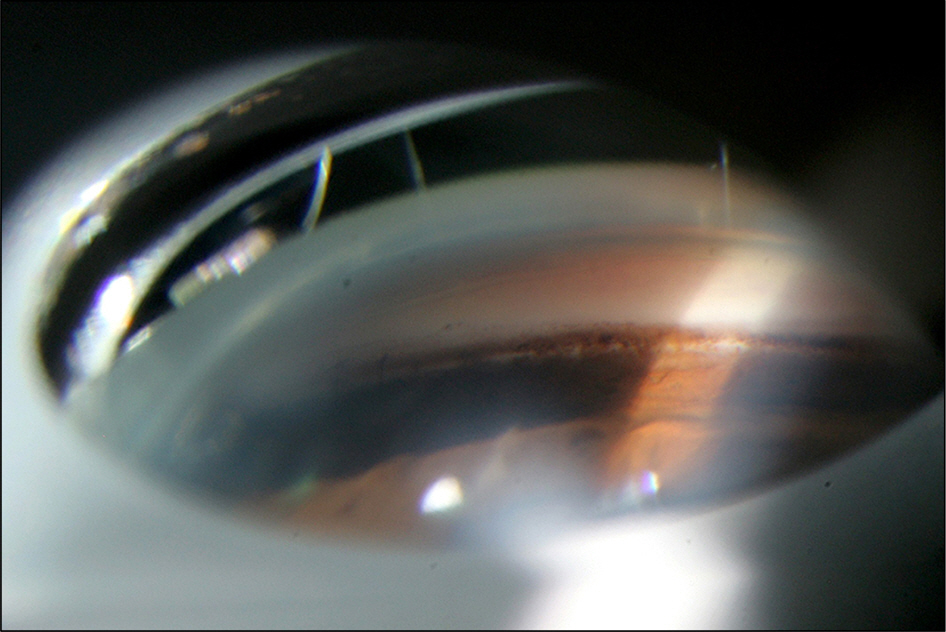
Figure 2. Inferior gonioscopy photograph of the left eye shows that the angle is open, and that the dense uniform trabecular meshwork pigmentations are observed at the lower 120 degrees.

Figure 3. Fundus photographss of the patient do notes not show glaucomatous cupping or retinal nerve fiber layer defect. Multiple hard exudates are observed in the both retinas, which are regarded as senile degenerative changes.
Reference
-
References
1. Sturrock GD, Tripathi RC.Pigmented lens striae. Br J Ophthalmol. 1976; 60:287–93.
Article2. Niyadurupola N, Broadway DC.Pigment dispersion syndrome and pigmentary glaucoma–a major review. Clin Experiment Ophthalmol. 2008; 36:868–82.3. Roberts DK, Winters JE, Castells DD. . Pigmented striae of the anterior lens capsule and age-associated pigment dispersion of var-iable degree in a group of older African-Americans: an age, race, and gender matched study. Int Ophthalmol. 2001; 24:313–22.
Article4. Roberts DK, Winters JE, Castells DD. . A cross-sectional study of Krukenberg spindles and pigmented lens striae in a predom-inately black population: two highly associated clinical signs of an-terior segment pigment dispersal. J Glaucoma. 2005; 14:57–63.5. Roberts DK, Wilensky J.Long anterior lens zonules. Clin Experiment Ophthalmol. 2012; 40:764–6.
Article6. Moroi SE, Lark KK, Sieving PA. . Long anterior zonules and pigment dispersion. Am J Ophthalmol. 2003; 136:1176–8.
Article7. Qing G, Wang N, Tang X. . Clinical characteristics of pigment dispersion syndrome in Chinese patients. Eye (Lond). 2009; 23:1641–6.
Article8. Ritch R.Exfoliation syndrome. Curr Opin Ophthalmol. 2001; 12:124–30.
Article9. Ritch R, Schlötzer-Schrehardt U.Exfoliation syndrome. Surv Ophthalmol. 2001; 45:265–315.
Article10. Hong C, Han SH, Sohn YH.A case of pigmentary glaucoma. J Korean Ophthalmol Soc. 1983; 24:435–9.11. Drolsum L, Ringvold A, Nicolaissen B.Cataract and glaucoma surgery in pseudoexfoliation syndrome: a review. Acta Ophthalmol Scand. 2007; 85:810–21.
Article12. Choi J, Park KH.Clinical characteristics of Korean patients with pseudoexfoliation syndrome. J Korean Ophthalmol Soc. 2006; 47:577–86.13. Subrayan V, Morris B, Armbrecht AM. . Long anterior lens zonules in late-onset retinal degeneration (L-ORD). Am J Ophthalmol. 2005; 140:1127–9.
Article14. Milam AH, Curcio CA, Cideciyan AV. . Dominant late-onset retinal degeneration with regional variation of sub-retinal pigment epithelium deposits, retinal function, and photoreceptor degeneration. Ophthalmology. 2000; 107:2256–66.
Article
- Full Text Links
-
- Actions
-
Cited
- CITED
-
- Close
- Share
-
- Similar articles
-
- Lens Dislocation during Extracapsular Cataract Extraction in Exfoliation Syndrome: A Case Report
- Pigment Dispersion Syndrome and Reverse Pupillary Block after Implantable Collamer Lens with Central Hole Implantation
- The Lens Dislocation into the Anterior Chamber in a case of Marfan's syndrome
- Clinical Result of One-Piece Aspheric Intraocular Lens in Mild Zonule-Weakness
- A Case of Anterior Lenticonus in Alport's Syndrome
