Pigment Dispersion Syndrome and Reverse Pupillary Block after Implantable Collamer Lens with Central Hole Implantation
- Affiliations
-
- 1HanGil Eye Hospital, Incheon, Korea. artemismj@hanmail.net
- KMID: 2355426
- DOI: http://doi.org/10.3341/jkos.2016.57.10.1661
Abstract
- PURPOSE
To report a case of pigment dispersion syndrome and reverse pupillary block secondary to the implantation of implantable collamer lens (ICL) with a central hole (AQUA ICL®) that was treated with ICL removal and laser peripheral iridotomy (LPI).
CASE SUMMARY
A 29-year-old woman with myopia in both eyes underwent implantation of AQUA ICL®. Four weeks postoperatively, the intraocular pressure (IOP) increased to 34 mm Hg and the patient showed pigment dispersion syndrome in both eyes. Since the IOP did not reduce with the maximum tolerable medical therapy, the ICLs were removed 8 weeks after implantation. The pigment dispersion subsided and IOP reduced shortly after ICL removal. However, 4 weeks after removal of ICL, posterior iris bowing and reverse pupillary block occurred in the right eye and the IOP increased to 46 mm Hg. LPI was performed in the right eye, and the reverse pupillary block was dissolved after a reduction in pigment dispersion. The IOP subsequently normalized to 13 mm Hg. Two weeks later, prophylactic LPI was performed in the left eye. Four weeks after prophylactic LPI, selective laser trabeculoplasty was performed on both eyes. As a result, the IOP was 11 mm Hg in the right eye and 12 mm Hg in the left eye after 4 weeks of treatment with topical IOP-lowering medications.
CONCLUSIONS
The present case indicates that implantation of ICL with a central hole can lead to early postoperative pigment dispersion syndrome. When this condition persists and is accompanied by reverse pupillary block after ICL removal, LPI can be partially effective.
Keyword
Figure
-
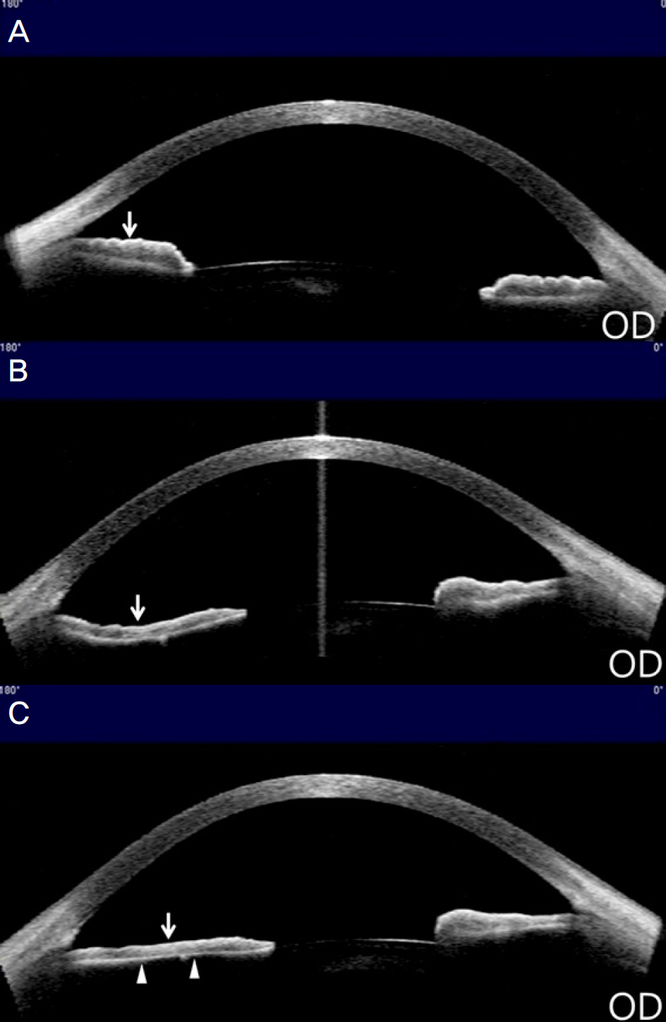
Figure 1. Changes in the iris contour of patient's right eye. Anterior segment optical coherence tomography images showing that the iris of the right eye was planar (white arrow) before implantation of AQUA ICL® (A) and became concave (white arrow) because of posterior iris bowing after AQUA ICL® removal (B). The iris became planar (white arrow) after laser peripheral iridotomy. Thinning of iris stroma (white arrowheads) was observed (C). OD = oculus dexter.
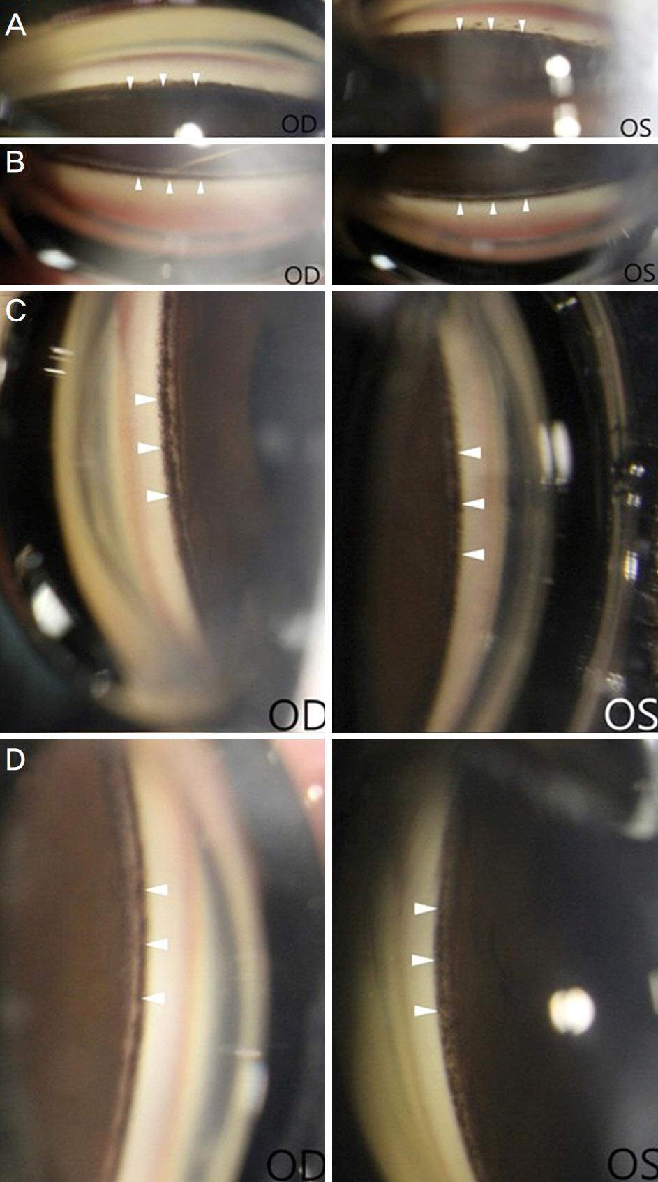
Figure 2. Gonioscopy showing open angles with 3 degrees of pigment deposition on the trabecular meshwork (white arrowheads) in the right eye and 2 degrees of pigment deposition in the left eye at 4 weeks after laser peripheral iridotomy (LPI) on the right eye (2 weeks after LPI on the left eye). (A) Inferior, (B) superior, (C) nasal, and (D) temporal side of the gonioscopic view. OD = oculus dexter; OS = oculus sinister.
Reference
-
References
1. Sanders DR, Doney K, Poco M. ICL in Treatment of Myopia Study Group. United States Food and Drug Administration clinical trial of the Implantable Collamer Lens (ICL) for moderate to high abdominal: three-year follow-up. Ophthalmology. 2004; 111:1683–92.2. Fernandes P, González-Méijome JM, Madrid-Costa D, et al. Implantable collamer posterior chamber intraocular lenses: a abdominal of potential complications. J Refract Surg. 2011; 27:765–76.3. Sánchez-Galeana CA, Zadok D, Montes M, et al. Refractory abdominal pressure increase after phakic posterior chamber intraocular lens implantation. Am J Ophthalmol. 2002; 134:121–3.4. Pak KY, Kim HS, Lee JW. A case of pigmentary glaucoma after posterior chamber phakic intraocular lens implantation. J Korean Ophthalmol Soc. 2013; 54:994–9.
Article5. Brandt JD, Mockovak ME, Chayet A. Pigmentary dispersion abdominal induced by a posterior chamber phakic refractive lens. Am J Ophthalmol. 2001; 131:260–3.6. Abela-Formanek C, Kruger AJ, Dejaco-Ruhswurm I, et al. Gonioscopic changes after implantation of a posterior chamber lens in phakic myopic eyes. J Cataract Refract Surg. 2001; 27:1919–25.
Article7. Son GS, Kim JW, Lim TH, et al. Comparison of clinical outcomes in implantable collamer lens implantation between AQUA ICL(R) and conventional ICL. J Korean Ophthalmol Soc. 2015; 56:1316–23.8. Lisa C, Naveiras M, Alfonso-Bartolozzi B, et al. Posterior chamber collagen copolymer phakic intraocular lens with a central hole to correct myopia: one-year follow-up. J Cataract Refract Surg. 2015; 41:1153–9.
Article9. Campbell DG. Pigmentary dispersion and glaucoma. A new theory. Arch Ophthalmol. 1979; 97:1667–72.10. Karickhoff JR. Pigmentary dispersion syndrome and pigmentary glaucoma: a new mechanism concept, a new treatment, and a new technique. Ophthalmic Surg. 1992; 23:269–77.
Article11. Alfonso JF, Lisa C, Abdelhamid A, et al. Three-year follow-up of subjective vault following myopic implantable collamer lens implantation. Graefes Arch Clin Exp Ophthalmol. 2010; 248:1827–35.
Article12. Chung TY, Park SC, Lee MO, et al. Changes in iridocorneal angle structure and trabecular pigmentation with STAAR implantable collamer lens during 2 years. J Refract Surg. 2009; 25:251–8.
Article13. Kawamorita T, Uozato H, Shimizu K. Fluid dynamics simulation of aqueous humour in a posterior-chamber phakic intraocular lens with a central perforation. Graefes Arch Clin Exp Ophthalmol. 2012; 250:935–9.
Article14. Scott A, Kotecha A, Bunce C, et al. YAG laser peripheral iridotomy for the prevention of pigment dispersion glaucoma a abdominal, randomized, controlled trial. Ophthalmology. 2011; 118:468–73.
- Full Text Links
-
- Actions
-
Cited
- CITED
-
- Close
- Share
-
- Similar articles
-
- Pupillary Block Following Phacoemulsification with Foldable Intraocular Lens Implantation in Adults
- Short-term Clinical Outcomes of Implantable Collamer Lens Implantation with Simultaneous Full Thickness Astigmatic Keratotomy
- Long-term Clinical Outcomes of Implantable Collamer Lens
- Axial Length Change after Implantable Collamer Lens Implantation
- IOP and Gonioscopic Changes after Implantable Contact Lens Implantation in Myopic Eyes
