J Korean Ophthalmol Soc.
2012 Jun;53(6):775-780.
Choice of One-Piece Intraocular Lens Power and Changes of Anterior Chamber in Sulcus Implantation due to Posterior Capsular Rupture during Cataract Surgery
- Affiliations
-
- 1Department of Ophthalmology and The Institute of Vision Research, Yonsei University College of Medicine, Seoul, Korea.
- 2Siloam Eye Hospital, Seoul, Korea. jhkim32@hanmail.net
- 3Jeil Eye Clinic, Suwon, Korea.
Abstract
- PURPOSE
To evaluate the appropriate correction of predicted intraocular lens (IOL) power when sulcus implantation due to posterior capsular rupture was performed and to compare the anterior chamber angle of sulcus-implanted eyes with in-the-bag implanted eyes using anterior segment optical coherence tomography (Visante OCT).
METHODS
Fifty-two eyes of 52 patients who had IOL implantation in the sulcus due to posterior capsular rupture during cataract surgery were retrospectively reviewed. A 0.5 lower diopter IOL than predicted IOL power using SRK-T formula was chosen. The difference between the predicted refractive error and the spherical equivalence at the 3-month time-point after sulcus implantation were evaluated. Among 52 patients, 16 patients whose fellow eye had an in-the-bag IOL implantation underwent Visante OCT and the anterior angle of affected eyes and normal fellow eyes were compared.
RESULTS
The difference between the predicted refractive error using SRK-T formula and the spherical equivalence after sulcus implantation was a myopic shift of 0.597 +/- 0.879 diopter. The mean iridocorneal angles, angle opening distance (AOD), trabecular iris surface area (TISA) at 500 um and 750 um for both nasal and temporal sides in sulcus implantation were all significantly lower than in-the-bag implantation.
CONCLUSIONS
A choice of a 0.5 lower diopter power IOL than the power for in-the-bag implantation may be considered because of greater myopic shift than predicted refractive error when sulcus implantation due to posterior rupture is performed. Anterior chamber angle in sulcus IOL implantation was shallower than in-the-bag IOL implantation.
Keyword
MeSH Terms
Figure
-
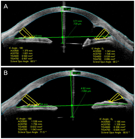
Figure 1 Anterior segment optical coherence tomography (Visante OCT) images show implanted intraocular lens (IOL) in the sulcus (A) and in the bag (B). IOL is placed more anterior than predicted position when implanted in the sulcus, which leads to myopic shift.
Reference
-
1. Jaycock P, Johnston RL, Taylor H, et al. The Cataract National Dataset electronic multi-centre audit of 55,567 operations: updating benchmark standards of care in the United Kingdom and internationally. Eye (Lond). 2009. 23:38–49.2. Zaidi FH, Corbett MC, Burton BJ, Bloom PA. Raising the benchmark for the 21st century--the 1000 cataract operations audit and survey: outcomes, consultant-supervised training and sourcing NHS choice. Br J Ophthalmol. 2007. 91:731–736.3. Bayramlar H, Hepsen IF, Yilmaz H. Myopic shift from the predicted refraction after sulcus fixation of PMMA posterior chamber intraocular lenses. Can J Ophthalmol. 2006. 41:78–82.4. Spokes DM, Norris JH, Ball JL. Refinement of lens power selection for sulcus placement of intraocular lens. J Cataract Refract Surg. 2010. 36:1436–1437.5. Suto C, Hori S, Fukuyama E, Akura J. Adjusting intraocular lens power for sulcus fixation. J Cataract Refract Surg. 2003. 29:1913–1917.6. Shin CJ, Lee JE, Kim JY, Tchah HW. Changes in anterior chamber depth and angle after phacoemulsification measured by anterior segment optical coherence tomography. J Korean Ophthalmol Soc. 2010. 51:353–358.7. Kucumen RB, Yenerel NM, Gorgun E, et al. Anterior segment optical coherence tomography measurement of anterior chamber depth and angle changes after phacoemulsification and intraocular lens implantation. J Cataract Refract Surg. 2008. 34:1694–1698.8. Nolan WP, See JL, Aung T, et al. Changes in angle configuration after phacoemulsification measured by anterior segment optical coherence tomography. J Glaucoma. 2008. 17:455–459.9. Knox Cartwright NE, Aristodemou P, Sparrow JM, Johnston RL. Adjustment of intraocular lens power for sulcus implantation. J Cataract Refract Surg. 2011. 37:798–799.10. Maeng HS, Ryu EH, Chung TY, Chung ES. Effects of anterior chamber depth and axial length on refractive error after intraocular lens implantation. J Korean Ophthalmol Soc. 2010. 51:195–202.11. Vasavada AR, Raj SM, Karve S, et al. Retrospective ultrasound biomicroscopic analysis of single-piece sulcus-fixated acrylic intraocular lenses. J Cataract Refract Surg. 2010. 36:771–777.12. Taskapili M, Gulkilik G, Kocabora MS, et al. Comparison of sulcus implantation of single-piece hydrophilic foldable acrylic and polymethylmethacrylate intraocular lenses in eyes with posterior capsule tear during phacoemulsification surgery. Eur J Ophthalmol. 2007. 17:595–600.13. Chang DF, Masket S, Miller KM, et al. Complications of sulcus placement of single-piece acrylic intraocular lenses: recommendations for backup IOL implantation following posterior capsule rupture. J Cataract Refract Surg. 2009. 35:1445–1458.14. LeBoyer RM, Werner L, Snyder ME, et al. Acute haptic-induced ciliary sulcus irritation associated with single-piece AcrySof intraocular lenses. J Cataract Refract Surg. 2005. 31:1421–1427.15. Micheli T, Cheung LM, Sharma S, et al. Acute haptic-induced pigmentary glaucoma with an AcrySof intraocular lens. J Cataract Refract Surg. 2002. 28:1869–1872.
- Full Text Links
-
- Actions
-
Cited
- CITED
-
- Close
- Share
-
- Similar articles
-
- Posterior Capsular Rupture in Planned ECCE for Posterior Chamber Intraocular Lens Implantation
- Intraocular Pressure Changes in the Uneventful Extra-capsular Cataract Extraction and Extra-capsular Cataract Extraction with Vitreous Loss followed by Anterior Chamber Lens Implantation
- Implantation of Posterior Chamber Lens in the Absenee of Posterior Capsular Support
- Intraocular Pressure after Extracapsular Cataract Extraction with Implantation of Posterior Chamber Lenses
- Intraocular pressure after extracapsular cataract extraction with implantation of posterior chamber lenses.
