J Korean Ophthalmol Soc.
2008 Oct;49(10):1634-1640.
Changes in RNFL Thickness According to the Myopia in Patients with Glaucoma and Ocular Hypertension
- Affiliations
-
- 1Department of Ophthalmology, Korea University College of Medicine, Seoul, Korea. yongykim@mail.korea.ac.kr
Abstract
- PURPOSE
To evaluate the changes in retinal nerve fiber layer (RNFL) thickness according to the degree of myopia in patients with glaucoma and ocular hypertension.
METHODS
Ninety-eight patients (165 eyes) diagnosed with glaucoma or ocular hypertension underwent optical coherence tomography (OCT) and scanning laser polarimetry using variable corneal compensation (GDx-VCC) to analyze the correlation between the degree of myopia and the thickness of the RNFL. A partial correlation coefficient analysis was performed to adjust for various factors such as age, laterality, intraocular pressure, and the mean deviation from visual field test, which can influence the RNFL thickness.
RESULTS
The average, nasal, superior, and inferior sectorial RNFL thicknesses measured by OCT significantly decreased with increasing myopia (p<0.05). However, RNFL thickness measured by GDx-VCC was not significantly correlated with the degree of myopia.
CONCLUSIONS
The RNFL thickness measured by OCT decreased with increasing myopia in eyes with glaucoma and ocular hypertension.
Keyword
MeSH Terms
Figure
-
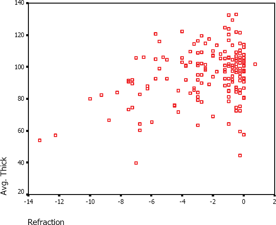
Figure 1. Scattergram of the average retinal nerve fiber layer (RNFL) thickness obtained by optical coherence tomography (y) and the degree of myopia (x). As myopia increased, there was a significant decrease in the average RNFL Thickness obtained by the Stratus OCT ( p=0.000, r=0.323).
Reference
-
References
1. Quigley HA, Katz J, Derrick RJ. . An evaluation of optic disc and nerve fiber layer examinations in monitoring progression of early glaucoma damage. Ophthalmology. 1992; 99:19–28.
Article2. Sommer HA, Quigley HA, Robin AL. . Evaluation of nerve fiber layer assessment. Arch Ophthalmol. 1984; 102:1766–71.
Article3. Quigley HA, Addicks EM, Green WR. Optic nerve damage in human glaucoma. Arch Ophthalmol. 1982; 100:135–46.
Article4. Hyung SM, Kim DM, Hong C, Youn DH. Optic Disc of the Myopic Eye: Relationship between Refractive Errors and Morphometric Characteristics. Korean J Ophthalmol. 1992; 6:32–5.
Article5. Curtin BJ, Karlin DB. Axial length measurements and fundus changes of the myopic eye. Am J Ophthalmol. 1971; 1:42–53.
Article6. Yanoff M, Fine BS. Ocular pathology: A text and atlas. 3rd ed. Philadelphia: JB Lippincott;1989. p. 408.7. Tomlinson A, Philips CI. Ratio of optic cup to optic disc in relation to axial length of eyeball and refraction. Br J Ophthalmol. 1969; 53:765.
Article8. Daubs JG, Crick RP. Effect of refractive error on the risk of ocular hypertension and open angle glaucoma. Trans Ophthal Soc U K. 1981; 101:121.9. Perkins ES, Phelps CD. Open angle glaucoma, ocular hypertension, low-tension glaucoma, and refraction. Arch Ophthalmol. 1982; 100:1464.
Article10. Phelps CD. Effects of myopia on prognosis in treated primary open-angle glaucoma. Am J Ophthalmol. 1982; 93:622.11. Curtin BJ. The Myopias. Philadelphia: Harper & Row;1985. p. 159–269.12. Jonas JB, Gusek GC, Naumann GOH. Optic disk morphometry in high myopia. Graefe’s Arch Clin Exp Ophthalmol. 1988; 226:587.
Article13. Johnstone MA. Ritch R, Shields MB, Krupin T, editors. Primary open-angle glaucoma. The Glaucomas. 1st ed. St. Louis: C. V. Mosby;1989. v.2:p. chap. 35.14. Huang D, Swanson EA, Lin CP. . Optical coherence tomography. Science. 1991; 254:1178–81.
Article15. Hee MR, Izatt JA, Swanson EA. . Optical coherence tomography of the human retina. Arch Ophthalmol. 1995; 113:325–32.
Article16. Budenz DL, Michael A, Chang RT. . Sensitivity and Specificity of the Stratus OCT for perimetric Glaucoma. Ophthalmology. 2005; 112:3–9.17. Malgorzata M, Bakunowice-Lazarczuk A, Sredzmskn-Kita D. Use of optical coherence tomography in myopia. J Pediatr Ophthalmol Strabismus. 2004; 41:159–62.
Article18. Weinreb RN. Evaluating the retinal nerve fiber layer in glaucoma with scanning laser polarimetry. Arch Ophthalmol. 1999; 117:1403–6.
Article19. Morgan JE, Waldock A, Jeffery G, Cowey A. Retinal nerve fiber layer polarimetry: histological and clinical comparison. Br J Ophthalmol. 1998; 82:684–90.20. Niessen AG, Van Den Berg TJ, Langerhorst CT, Greeve EL. Retinal nerve fiber layer assessment by scanning laser polarimetry and standarded photography. Am J Ophthalmol. 1996; 121:484–93.21. Tjon-Fo-Sang MJ, Lemij HG. The sensitivity and specificity of nerve fiber measurements in glaucoma as determined with scanning laser polarimetry. Am J Ophthalmol. 1997; 123:62–9.22. Zhou Q, Weinreb RN. Individualized compensation of anterior segment birefringence during scanning laser polarimetry. Invest Ophthalmol Vis Sci. 2002; 43:2221–8.23. Weinreb RN, Bowd C, Zangwill LM. Glaucoma detection using scanning laser polarimetry with variable corneal polarization compensation. Arch Ophthalmol. 2003; 121:218–24.
Article24. Greenfield DS, Knighton RW, Huang XR. Effect of corneal polarization axis on assessment of retinal nerve fiber layer thickness by scanning laser polarimetry. Am J Ophthalmol. 2000; 129:715–22.
Article25. Park MH, Hwang HS, Moon JI. Diagnosis of Glaucoma in the cases of the opposed Results of GDx and OCT. J Korean Ophthalmol Soc. 2005; 46:2010–5.26. Choi SW, Lee SJ. Thickness Changes in the Fovea and Peripapillary Retinal Nerve Fiber Layer Depend on the Degree of Myopia. Korean J Ophthalmol. 2006; 20:215–9.
Article27. Bowd C, Medeiros FA, Weinreb RN, Zangwill LM. The Effect of Atypical Birefringence Patterns on Glaucoma Detection Using Scanning Laser Polarimetry with Variable Corneal Compensation. Invest Ophthalmol Vis Sci. 2007; 48:223–7.
Article28. Leung CK, Mohamed S, Leung KS. . Retinal nerve Fiber Layer Measurements in Myopia: An Optical Coherence Tomography Study. Invest Ophthalmol Vis Sci. 2006; 47:5171–6.
Article29. Ozdek SC, Onol M, Gürelik G, Hasanreisoglu B. Scanning laser polarimetry in normal subjects and patients with myopia. Br J Ophthalmol. 2000; 84:264–7.
Article30. Kremmer S, Zadow T, Steuhl KP, Selbach JM. Scanning laser polarimetry in myopic and hyperopic subjects. Graefes Arch Clin Exp Ophthalmol. 2004; 242:489–94.
Article31. Salchow DJ, Oleynikov YS, Chiang MF. . Retinal nerve fiber layer thickness in normal children measured with optical coherence tomography. Ophthalmology. 2006; 113:786–91.
Article32. Bowd C, Zangwill LM, Blumenthal EZ. . Imaging of the optic disc and retinal nerve fiber layer: the effects of age, optic disc area, refractive error, and gender. J Opt Soc Am A Opt Image Sci Vis. 2002; 19:197–207.
Article33. Hoh ST, Lim MC, Seah SK. . Peripapillary retinal nerve fiber layer thickness variations with myopia. Ophthalmology. 2006; 113:773–7.
Article34. Bozkurt B, Irkec M, Gedik S. Effect of peripapillary chorioretinal atrophy on GDx parameters in patients with degenerative myopia. Clin Exp Ophthalmol. 2002; 30:411–4.
Article35. Mitchell P, Hourihan F, Sandbach J, Wang JJ. The relationship between glaucoma and myopia: the Blue Mountains Eye Study. Ophthalmology. 1999; 106:2010–15.36. Grodum K, Heijl A, Bengtsson B. Refractive error and glaucoma. Acta Ophthalmol Scand. 2001; 79:560–6.
- Full Text Links
-
- Actions
-
Cited
- CITED
-
- Close
- Share
-
- Similar articles
-
- The Structure-function Relationships between Two Different Optical Coherence Tomography in Patients with High Myopic Glaucoma
- Thickness Changes in the Fovea and Peripapillary Retinal Nerve Fiber Layer Depend on the Degree of Myopia
- Factors Affecting Post-Cataract OCT Parameters in Open-Angle and Angle-Closure Glaucoma Eyes: Optic Disc Head Analysis
- The Relationship between Open Angle Glaucoma and Refractive Errors
- Correlation Between Macular, Retinal Nerve Fiber Layer Thickness, and Visual Field in Open Angle Glaucoma
