Thickness Changes in the Fovea and Peripapillary Retinal Nerve Fiber Layer Depend on the Degree of Myopia
- Affiliations
-
- 1Department of Ophthalmology, Wonju Christian Hospital. Yonsei University, Wonju College of Medicine, Wonju, Korea. eyesj@yonsei.ac.kr
- KMID: 754585
- DOI: http://doi.org/10.3341/kjo.2006.20.4.215
Abstract
- PURPOSE: To investigate changes in the thickness of the fovea and peripapillary RNFL associated with myopia. METHODS: Sixty-five Korean adults (for a total of 130 eyes) between 23 and 26 years of age were selected as test subjects. Thirty-eight test subjects were male, and 27 were female. Subjects with glaucoma or other identified ocular diseases were excluded. Patients whose manifest refraction measurement values ranged between 0 to -2D were classified as group one (emmetropia and low myopia), those between -2 to -5D were classified as group two (moderate myopia), and those more than -5D were classified as group three (high myopia). Using the OCT, the thickness of the fovea and peripapillary RNFL were measured for every subject. RESULTS: The thicknesses of the fovea for each of three groups were 142.16+/-8.99 micrometer in group one (45 eyes), 153.58+/-17.63 micrometer in group two (43 eyes) and 158.86+/-11.93 micrometer in group three (28 eyes). The data showed significant differences in fovea thickness between the groups. The average thicknesses of the peripapillary RNFL for each of three groups were 113.29+/-10.80 micrometer in group one, 103.85+/-14.48 micrometer in group two and 100.74+/-9.15 micrometer in group three. A statistically significant difference was found between group one and the other groups (p<0.05). CONCLUSIONS: As the level of myopia increased, the thickness of the fovea also increased, while the thickness of the peripapillary RNFL decreased. Therefore, when interpreting OCT results in the clinic, careful consideration should be given to various changes associated with myopia.
Keyword
MeSH Terms
Figure
-
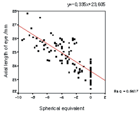
Fig. 1 Relationship between axial length and myopia.

Fig. 2 Relationship between foveal thickness and myopia.

Fig. 3 Relationship between RNFL-average thickness and myopia.
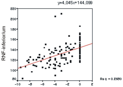
Fig. 4 Relationship between RNFL-inferior thickness and myopia.

Fig. 5 Relationship between RNFL-superior thickness and myopia.
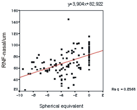
Fig. 6 Relationship between RNFL-nasal thickness and myopia.
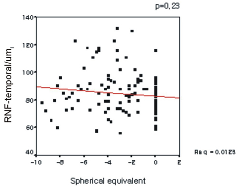
Fig. 7 Relationship between RNFL-temporal thickness and myopia.
Cited by 7 articles
-
Changes in RNFL Thickness According to the Myopia in Patients with Glaucoma and Ocular Hypertension
Jung Wan Kim, Yong Yeon Kim
J Korean Ophthalmol Soc. 2008;49(10):1634-1640. doi: 10.3341/jkos.2008.49.10.1634.Quantitative Analysis of Retinal Nerve Fiber Layer Thickness Profile in Myopic Eyes
Tae Geun Song, Young Cheol Yoo, Ha Bum Lee
J Korean Ophthalmol Soc. 2009;50(12):1840-1846. doi: 10.3341/jkos.2009.50.12.1840.Use of Spectral-Domain Optical Coherence Tomography to Analyze Macular Thickness According to Refractive Error
Seung Hoon Kim, Joo Youn Park, Tae Kwann Park, Young-Hoon Ohn
J Korean Ophthalmol Soc. 2011;52(11):1286-1295. doi: 10.3341/jkos.2011.52.11.1286.Changes of Peripapillary Retinal Nerve Fiber Layer Thickness Profile According to Aging in Myopic Eyes
Eun Jin Bae, Young Cheol Yoo
J Korean Ophthalmol Soc. 2013;54(7):1066-1073. doi: 10.3341/jkos.2013.54.7.1066.Comparison of Diagnostic Power Among OCT Parameters According to Peripapillary Atrophy in High Myopic Glaucoma
Woo Jin Kim, Kyung Nam Kim, Chang Sik Kim
J Korean Ophthalmol Soc. 2013;54(12):1844-1855. doi: 10.3341/jkos.2013.54.12.1844.The Effects of Optic Disc Factors on Retinal Nerve Fiber Layer Thickness Measurement in Children
Jong-Hwa Jun, Se-Youp Lee
Korean J Ophthalmol. 2008;22(2):115-122. doi: 10.3341/kjo.2008.22.2.115.The Effects of Optic Disc Factors on Retinal Nerve Fiber Layer Thickness Measurement in Children
Jong-Hwa Jun, Se-Youp Lee
Korean J Ophthalmol. 2008;22(2):115-122. doi: 10.3341/kjo.2008.22.2.115.
Reference
-
1. Hammond CJ, Shnieder H, Gilbert CE, Spector TD. Genes and environment in refractive error : The twin eye study. Invest Ophthalmol Vis Sci. 2001. 42:1232–1236.2. Young FA. The nature and control of myopia. J Am Optom Assoc. 1977. 48:451–457.3. Mutti DO, Mitchell GL, Moeschberger ML, et al. Parental myopia, near work, school achievement, and children's refractive error. Invest Ophthalmol Vis Sci. 2002. 43:3633–3640.4. Lin LL, Shih YF, Hsiao CK, et al. Epidemiologic study of the prevalence and severity of myopia among schoolchildren in Taiwan in 2000. J Formos Med Assoc. 2001. 100:684–691.5. Wong TY, Foster PJ, Hee J, et al. Prevalence and risk factors for refractive errors in adult Chinese in Singapore. Invest Ophthalmol Vis Sci. 2000. 41:2486–2494.6. Au Eong KG, Tay TH, Lim MK. Education and myopia in 110,236 young Singaporean males. Singapore Med J. 1993. 34:489–492.7. Tay MT, Au Eong KG, Ng CY, Lim MK. Myopia and educational attainment in 421,116 young Singaporean males. Ann Acad Med Singapore. 1992. 21:785–791.8. Kang SH, Kim PS, Choi DG. Prevalence of Myopia in 19-year-old Korean Males : The Relationship between the Prevalence and Education or Urbanization. J Korean Ophthalmol Soc. 2004. 45:2082–2087.9. Curtin BJ, Karlin DB. Axial length measurements and fundus changes of the myopic eye. Am J Ophthalmol. 1971. 1:42–53.10. Yanoff M, Fine BS. Ocular pathology: A text and atlas. 1989. 3rd ed. JB Lippincott;408.11. Budenz DL, Michael A, Chang RT, et al. Senditivity and Specificity of the StratusOCT for perimetric Glaucoma. Ophthalmology. 2005. 112:3–9.12. Malgorzata M, Alina BL, Dorota SK. Use of optical coherence tomography in myopia. J Pediatr Ophthalmol Strabismus. 2004. 41:159–162.13. Kanai K, Abe T, Murayama K. Retinal thickness and changes with age. Nippon Ganka Gakkai Zasshi. 2002. 106:162–165.14. Asrani S, Zou S, d'Anna S, et al. Noninvasive mapping of the normal retinal thickness at the posterior pole. Ophthalmology. 1999. 106:269–273.15. Jung Hee-jin, Hyun Jae Hoon, Kim Young Il, et al. Normal macular thickness measured macular mapping of OCT3. J Korean Ophthalmol Soc. 2004. 45:962–968.16. Balazsi AG, Rootman J, Drance SM, et al. The effect of age on the nerve fiber population of the hyman optic nerve. Am J Ophthalmol. 1984. 97:760–766.17. Johnson BM, Miao M, et al. Age-related decline of human optic nerve axon populations. Age. 1987. 10:5–9.18. Gobel W, Hartmann F, Haigis W. Determination of retinal thickness in relation to the age and axial length using optical coherence tomography. Ophthalmologe. 2001. 98:157–162.19. Wong AC, Chan CW, Hui SP. Relationship of gender, body mass index, and axial length with central retinal thickness using optical coherence tomography. Eye. 2005. 19:292–297.20. Lim MC, Hoh ST, Foster PJ, et al. Use of optical coherence tomography to assess variations in macular retinal thickness in myopia. Invest Ophthalmol Vis Sci. 2005. 46:974–978.21. Park WS, Lee SH. Comparison between New Threshold Visual Field Strategy, SITA - standard and Full Threshold Strategy and Their Clinical Usefulness. J Korean Ophthalmol Soc. 2000. 41:1187–1191.22. Bengtsson B, Olsson J, Heijl A. A new generation of algorithms for computerized threshold perimetry, SITA. Acta Ophthalmol Scand. 1997. 75:368–375.23. Grossniklaus HE, Green WR. Pathologic findings in pathologic myopia. Retina. 1992. 12:127–133.24. Huang D, Swanson EA, Lin CP, et al. Optical coherence tomography. Science. 1991. 254:1178–1181.25. Kent C. Creating a "virtual biopsy": improvements in technology make it possible to "see" more internal tissue than ever before. Ophthalmology Management. 2002. 6:111–112.26. StratusOCT user manual. 2003. 4.27. Lee JH, Ahn CS, Lee DY. Qauntification of retinal nerve fiber layer thickness in the normal subjects using optical coherence tomography. J Korean Ophthalmol Soc. 1999. 40:2804–2815.28. Caprioli J, Ortiz-Colberg R, Miller JM, et al. Measurement of peripapillary nerve fiber layer contour in glaucoma. Am J Ophthalmol. 1989. 108:404–413.29. Lee DY, Yu SY, Kwak HW. Quantitative analysis of macular thickness with OCT map. J Korean Ophthalmol Soc. 2004. 45:1496–1502.
- Full Text Links
-
- Actions
-
Cited
- CITED
-
- Close
- Share
-
- Similar articles
-
- Thicknesses of the Fovea and Retinal Nerve Fiber Layer in Amblyopic and Normal Eyes in Children
- Quantitative Analysis of Retinal Nerve Fiber Layer Thickness Profile in Myopic Eyes
- Reproducibility of Retinal Nerve Fiber Layer Thickness Evaluation by Nerve Fiber Analyzer
- Biometry of Retinal Nerve Fiber Layer Thickness by NFA
- Changes in Macular Retinal Layers and Peripapillary Nerve Fiber Layer Thickness after 577-nm Pattern Scanning Laser in Patients with Diabetic Retinopathy
