J Korean Rheum Assoc.
2009 Dec;16(4):318-322.
A Case of the Lumbar Spine Involvement and Sacroiliitis in a Patient with Gout
- Affiliations
-
- 1Department of Rheumatology, Hospital for Rheumatic Disease, Hanyang University College of Medicine, Seoul, Korea. sungyk@hanyang.ac.kr
- 2Department of Radiology, Hanyang University College of Medicine, Seoul, Korea.
Abstract
- Although gout often initially affects the peripheral joints, gout may also involve the axial joints. The radiologic changes of axial gout are more common than are clinically recognized. According to a recent report, when the spine CT images of peripheral gout were reviewed for features of axial gout, there was about a 14% frequency of suspected axial gout. The vertebral level and the finding with the most common spinal gouty changes were L4 and lumbar facet joint erosions. We describe here the case of a 36-year-old gout patient with low back and right buttock pain and his lesions were unexpectedly diagnostic of lumbar facet joint arthritis and right sacroiliitis.
MeSH Terms
Figure
-
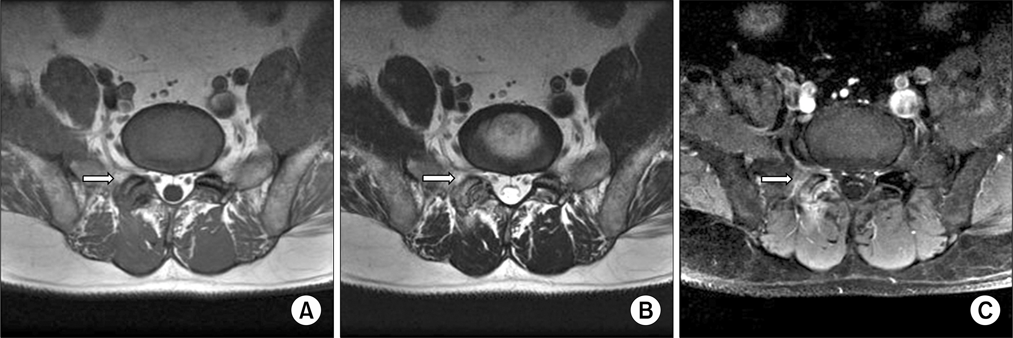
Fig. 1. The T1-weighted MRI shows low signal intensity on the right L5-S1 facet joint, bone, and soft tissue (A). T2-weighted MRI shows intermediate to high signal intensity on the right L5-S1 facet joint, bone, and soft tissue (B). The gadolinium enhanced T1-weighted MRI shows heterogenous enhancement of the right L5-S1 facet joint, bone, and soft tissue (C).
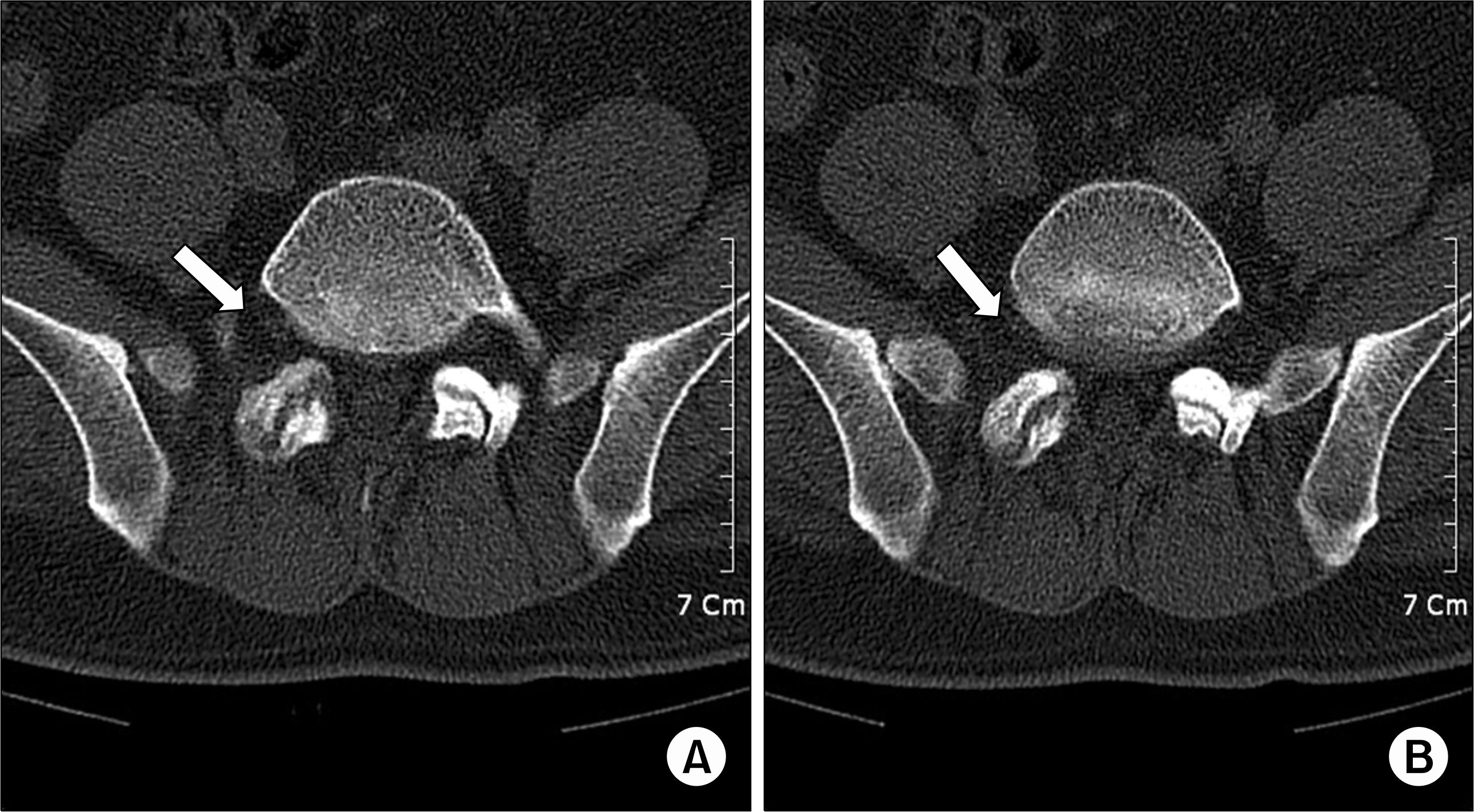
Fig. 2. The CT scan shows bony erosions in the right L5-S1 facet joint on two consecutive images (A, B).
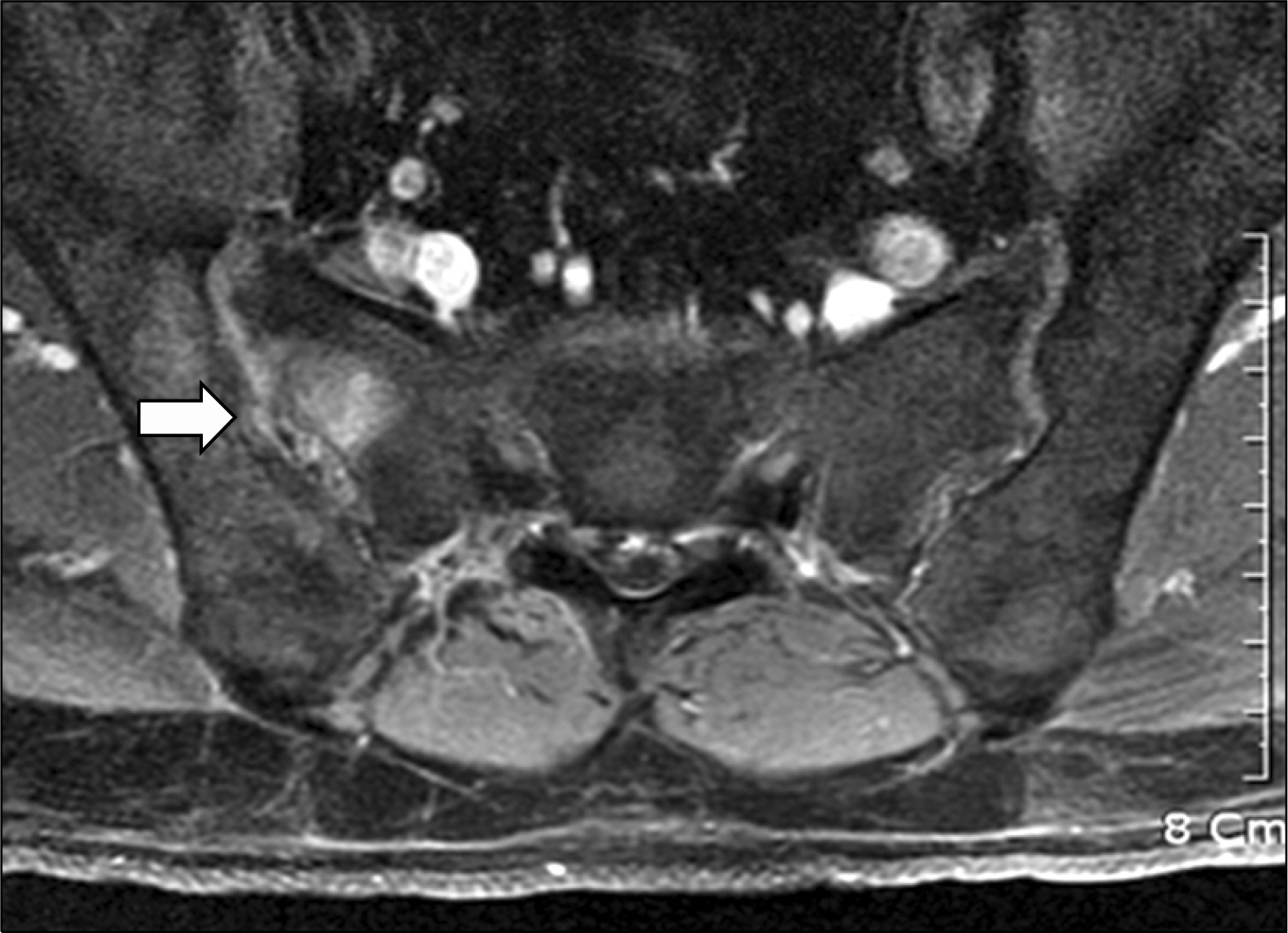
Fig. 3. The gadolinium enhanced T1-weighted MRI shows heterogenous enhancement of the right sacrum, SI joint and ilium.
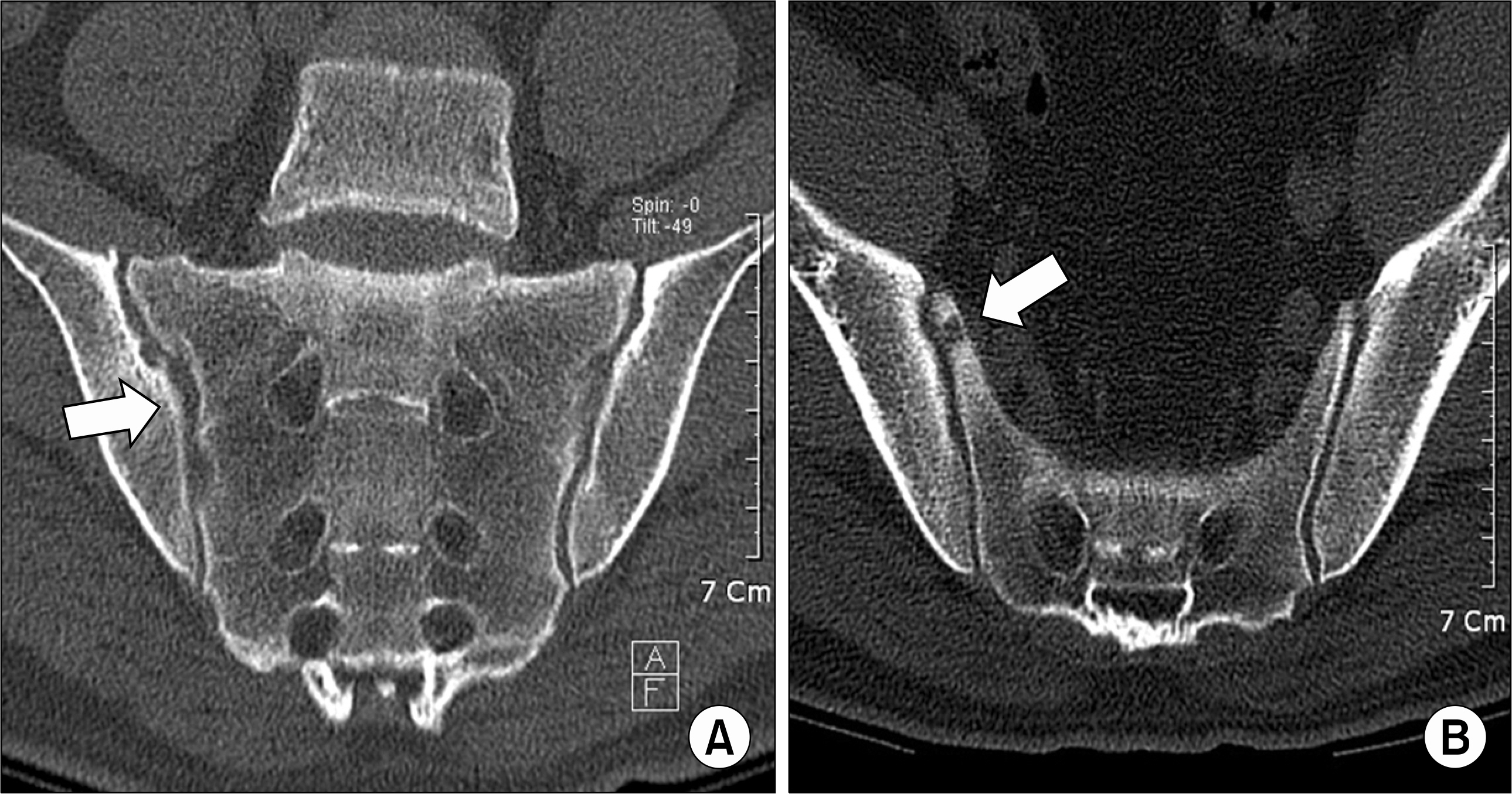
Fig. 4. The coronal reformatted CT image of the SI joints show multiple, well-marginated erosions along the articular surfaces of the right SI joint (A, B).
Reference
-
References
1. Grahame R, Scott JT. Clinical survey of 354 patients with gout. Ann Rheum Dis. 1970; 29:461–8.
Article2. Perkins P, Jones AC. Gout. Ann Rheum Dis. 1999; 58:611–7.
Article3. Schumacher HR. Crystal-induced arthritis: an overview. Am J Med. 1996; 100:S46–52.4. Kersley GD, Mandel L, Jeffrey MR. Gout; an unusual case with softening and subluxation of the first cervical vertebra and splenomegaly. Ann Rheum Dis. 1950; 9:282–304.5. Kaye PV, Dreyer MD. Spinal gout: an unusual clinical and cytological presentation. Cytopathology. 1999; 10:411–4.
Article6. van den Berge M, Vrugt B, Holt C, Smit CJ, Hoogenberg K. Gout as an unusual cause of pelvic pain. Ned Tijdschr Geneeskd. 2006; 150:151–4.7. Riddell CM, Elliott M, Cairns AP. An unusual "gouty" case of back pain and fever. J Rheumatol. 2008; 35:2076–7.8. Mantle B, Gross P, Lopez-Ben R, Alarcon GS. Hip pain as the presenting manifestation of acute gouty sacroiliitis. J Clin Rheumatol. 2001; 7:112–4.
Article9. Schlesinger N, Baker DG, Schumacher HR Jr. Serum urate during bouts of acute gouty arthritis. J Rheumatol. 1997; 24:2265–6.10. Konatalapalli RM, Demarco PJ, Jelinek JS, Murphey M, Gibson M, Jennings B, et al. Gout in the axial skeleton. J Rheumatol. 2009; 36:609–13.
Article11. Gerster JC, Landry M, Rappoport G, Rivier G, Du-voisin B, Schnyder P. Enthesopathy and tendinopathy in gout: computed tomographic assessment. Ann Rheum Dis. 1996; 55:921–3.
Article12. Gerster JC, Landry M, Rivier G. Computed tomographic imaging of subcutaneous gouty tophi. Clin Rheumatol. 1998; 17:62–4.
Article13. Oaks J, Quarfordt SD, Metcalfe JK. MR features of vertebral tophaceous gout. AJR Am J Roentgenol. 2006; 187:W658–9.
Article14. Hsu CY, Shih TT, Huang KM, Chen PQ, Sheu JJ, Li YW. Tophaceous gout of the spine: MR imaging features. Clin Radiol. 2002; 57:919–25.
Article15. King JC, Nicholas C. Gouty arthropathy of the lumbar spine: a case report and review of the literature. Spine. 1997; 22:2309–12.
- Full Text Links
-
- Actions
-
Cited
- CITED
-
- Close
- Share
-
- Similar articles
-
- Tophaceous Gout Involving the Whole Spine: An Unusual Case Report
- Complete Fusion of Three Lumbar Vertebral Bodies in Ankylosing Spondylitis
- Tophaceous Gout of the Spine Causing Neural Compression
- Chronic Tophaceous Gout in Multiple Spines: A Case Report and Literature Review
- Tophaceous Gout of the Lumbar Spine Mimicking Infectious Spondylodiscitis and Epidural Abscess
