Tophaceous Gout Involving the Whole Spine: An Unusual Case Report
- Affiliations
-
- 1Department of Radiology, Sanggye Paik Hospital, Inje University College of Medicine, Seoul, Korea. merita@paik.ac.kr
- KMID: 1439413
- DOI: http://doi.org/10.3348/jksr.2012.66.2.193
Abstract
- Gout is a relatively common, crystal deposition disease, in which monosodium urate crystals are deposited in joint and periarticular tissues of the extremities. Involvement of the spine is exceedingly rare. Most patients with spinal gout present with symptomatic spinal cord compression. Diffuse involvement of tophi deposition inside the spinal central canal has not been reported. We now present a case of chronic tophaceous gout with extensive spinal involvement that resulted in diffuse spinal cord compression and led to paraplegia.
Figure
-
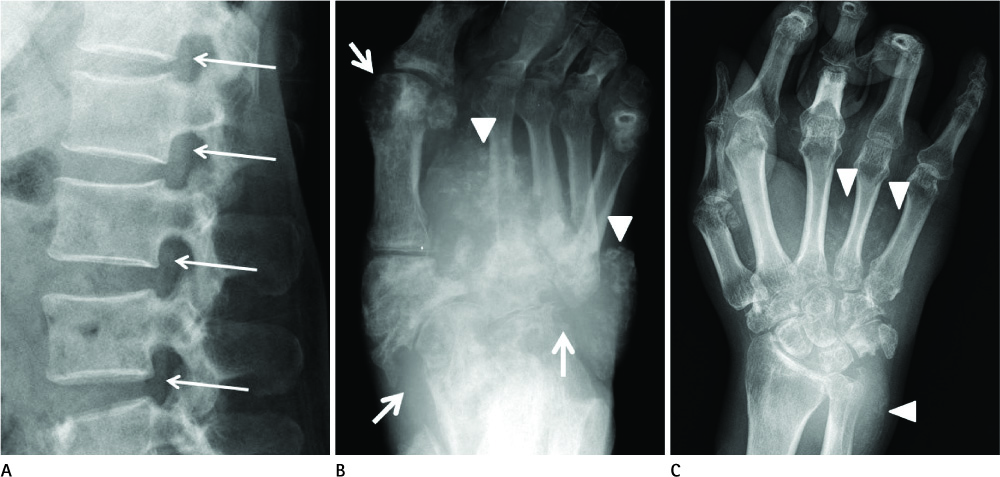
Fig. 1 Conventional radiographs of a 52-year-old man with paraplegia and arthralgia. A. On a lateral view of the lumbar spine, a hyperdense line is noted in the anterior portion of the central canal (arrows). B. A foot AP view shows multiple radioopaque depositions in soft tissue (arrowheads) and marginal bony erosions in the 1st metatarsophalangeal, tarsometatarsal, talonaivcular and calcaneocuboidal joints (arrows). Subluxation of the 2nd-5th metatarsal joints is seen. C. A apicoposterior view of wrist shows radioopaque tophi (arrowheads).
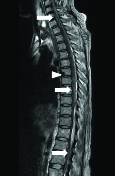
Fig. 2 A sagittal T2-weighted image of the whole spine demonstrating a long heterogeneous low signal band of tophi deposited in the anterior epidural space (arrows). A focal erosion is noted at the anterior margin of the T6 body (arrowhead). The spinal cord was compressed from C3 to T8.
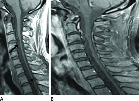
Fig. 3 MR images of cervical spine. A. T1-weighted sagittal MR images of cervical spine show heterogeneous intermediate (black arrow) to low (black arrowhead) signal intensity masses in the anterior epidural space, suggesting deposition of tophi. B. Fat-saturated T1-weighted sagittal images after gadolinium enhancement reveal no significant enhancement of tophi.
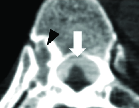
Fig. 4 Involvement of the costovertebral joint. Tophi are seen as hyperdense space-occupying lesions in the anterior epidural space (arrow) on an unenhanced axial CT image. Note the vertebral destruction around the right costovertebral joint due to deposition of tophi (black arrowhead).
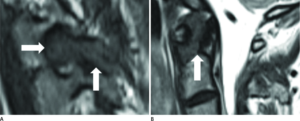
Fig. 5 Involvement of facet joints and the atlantodental joint. A. Axial and sagittal T1-weighted MR images show tophi in the facet joint (arrows), resulting in narrowing of the neural foramen. B. Deposition of tophi in the atlantodental interval on a sagittal T2-weighted image (arrow).
Cited by 1 articles
-
Chronic Tophaceous Gout in Multiple Spines: A Case Report and Literature Review
Kyoung Hwa Lee, Hyun Sun Woo, Mi Ryoung Seo, Hee Jung Ryu, Hyo Jin Choi, Han Joo Baek
J Rheum Dis. 2015;22(4):250-255. doi: 10.4078/jrd.2015.22.4.250.
Reference
-
1. Richette P, Bardin T. Gout. Lancet. 2010; 375:318–328.2. Ning TC, Keenan RT. Unusual clinical presentations of gout. Curr Opin Rheumatol. 2010; 22:181–187.3. Oaks J, Quarfordt SD, Metcalfe JK. MR features of vertebral tophaceous gout. AJR Am J Roentgenol. 2006; 187:W658–W659.4. Butteriss D, Soh C. A case of spinal cord compression of unknown cause. Br J Radiol. 2006; 79:775–777.5. Desai MA, Peterson JJ, Garner HW, Kransdorf MJ. Clinical utility of dual-energy CT for evaluation of tophaceous gout. Radiographics. 2011; 31:1365–1375. discussion 1376-13776. Paquette S, Lach B, Guiot B. Lumbar radiculopathy secondary to gouty tophi in the filum terminale in a patient without systemic gout: case report. Neurosurgery. 2000; 46:986–988.7. Yu JS, Chung C, Recht M, Dailiana T, Jurdi R. MR imaging of tophaceous gout. AJR Am J Roentgenol. 1997; 168:523–527.8. Hsu CY, Shih TT, Huang KM, Chen PQ, Sheu JJ, Li YW. Tophaceous gout of the spine: MR imaging features. Clin Radiol. 2002; 57:919–925.9. Bonaldi VM, Duong H, Starr MR, Sarazin L, Richardson J. Tophaceous gout of the lumbar spine mimicking an epidural abscess: MR features. AJNR Am J Neuroradiol. 1996; 17:1949–1952.10. Duprez TP, Malghem J, Vande Berg BC, Noel HM, Munting EA, Maldague BE. Gout in the cervical spine: MR pattern mimicking diskovertebral infection. AJNR Am J Neuroradiol. 1996; 17:151–153.
- Full Text Links
-
- Actions
-
Cited
- CITED
-
- Close
- Share
-
- Similar articles
-
- Tophaceous Gout Involving the Bipartitle Patella: A Case Report
- Tophaceous Gout in the Rotator Cuff with Impingement Syndrome: A Case Report
- Tophaceous Gout of the Lumbar Spine Mimicking Infectious Spondylodiscitis and Epidural Abscess
- Intratendinous Tophaceous Gout Mimicking Cellulitis after Achilles Tendon Repair
- Symptomatic Tophaceous Gout in the Bilateral Patellae
