Interaction between odontoblast and bio-calcium phosphate cement reinforced with chitosan
- Affiliations
-
- 1Department of Oral and Maxillofacial Surgery, School of Dentistry, Pusan National University, Yangsan, Korea. kuksjs@pusan.ac.kr
- 2Department of Oral Anatomy, School of Dentistry, Pusan National University, Yangsan, Korea.
Abstract
- PURPOSE
Calcium phosphate cement (CPC) is one of many useful materials for restoring tooth defects, periodontium and maxillofacial area. Chitosan is a biodegradable material that has been shown to promote the growth and differentiation of osteoblasts in culture. This study examined the interaction between odontoblasts and bio-calcium phosphate cement reinforced with chitosan.
MATERIALS AND METHODS
5x10(3) odontoblastic cells were seeded into each well. Various concentrations of bio-calcium phosphate cement reinforced with chitosan (10, 20, 50, 100, 200, 500 microg/ml, 1, 2, 4 mg/ml) were diluted and added to the wells. The well was incubated for 24 h, 48 h and 72 h. After incubation, the number of cells was assessed to determine the cell viability. A cytokinesis-block micronucleus assay and chromosomal aberration test were carried out to estimate the extent of chromosomal abnormalities. Microscopic photographs and RT-PCR were performed to examine the adhesion potential of bio-calcium phosphate cement reinforced with chitosan.
RESULTS
Bio-CPC-reinforced chitosan did not show significant cytotoxicity. The number of damaged chromosomes in the cells treated with Bio-CPC-reinforced chitosan was similar to that in the control cells. There was no significant increase in the number of chromosomal aberrations in the Bio-CPC reinforced chitosan exposed cells. Microscopic photographs and RT-PCR confirmed the adhesive potential of bio-CPC reinforced chitosan to odontoblasts.
CONCLUSION
Bio-CPC-reinforced chitosan did not affect the odontoblastic cell viability, and had no significant cytotoxic effect. Bio-CPC-reinforced chitosan showed adhesive potential to odontoblasts. These results are expected form the basis of future studies on the effectiveness of dental restorative materials in Bio-CPC reinforced with chitosan.
Keyword
MeSH Terms
Figure
-
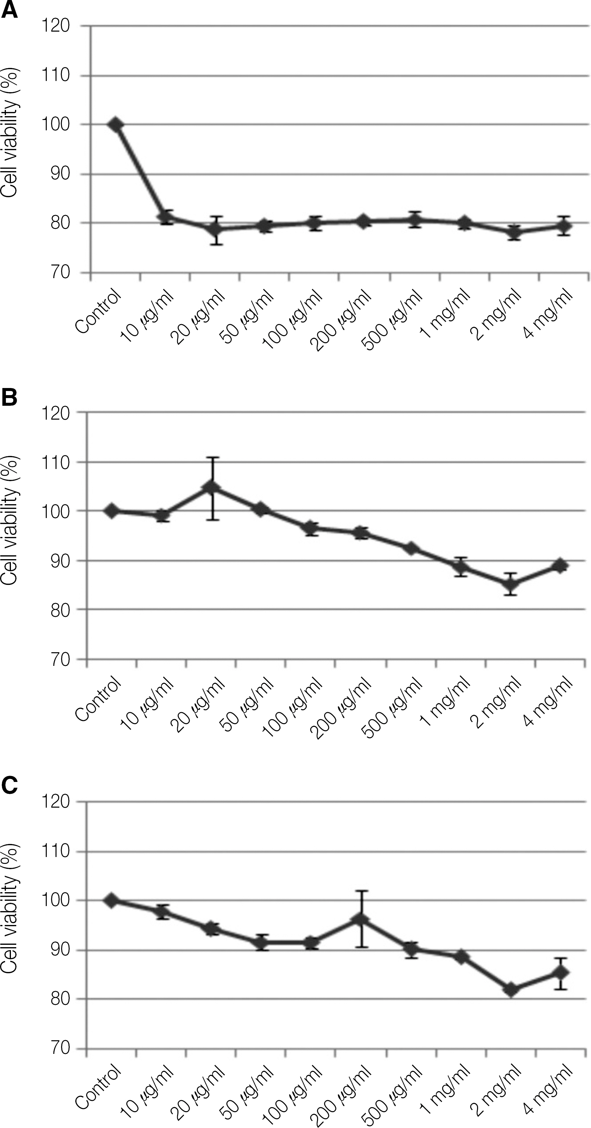
Fig. 1. The effects of cements on cell viability. Byung-Do Chun et al: Interaction between odontoblast and bio-calcium phosphate cement reinforced with chitosan. J Korean Assoc Oral Maxillofac Surg 2011
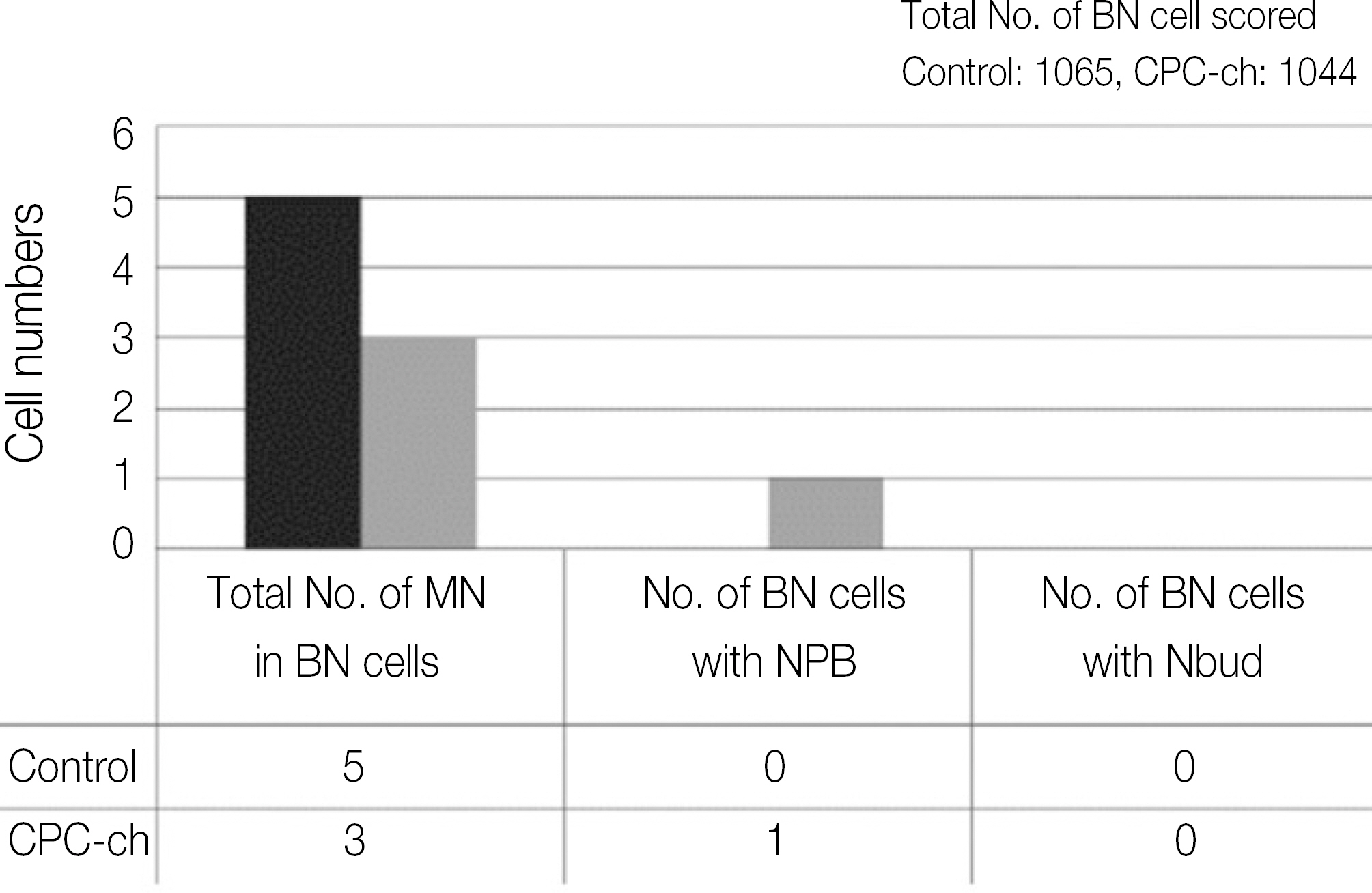
Fig. 2. Cytokinesis-block micronucleus assay. Byung-Do Chun et al: Interaction between odontoblast and bio-calcium phosphate cement reinforced with chitosan. J Korean Assoc Oral Maxillofac Surg 2011
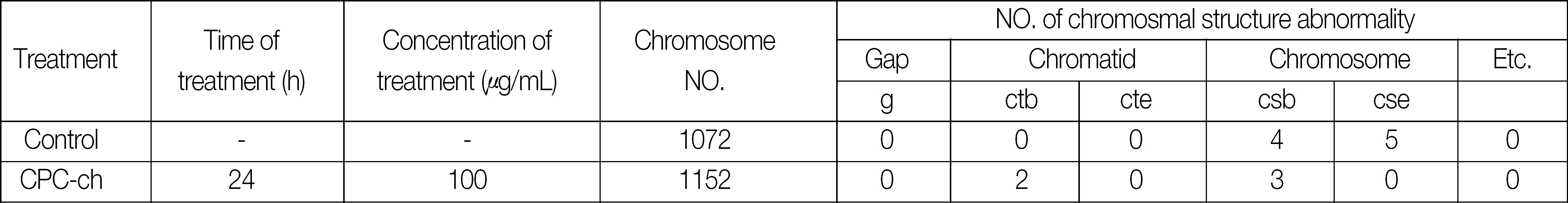
Fig. 3. Chromosomal abberration test.

Fig. 4. Microscopic photography of the cell adhesion. A: coated plate, B: non-coated plate, C: non-coated plate with CPC-ch. Byung-Do Chun et al: Interaction between odontoblast and bio-calcium phosphate cement reinforced with chitosan. J Korean Assoc Oral Maxillofac Surg 2011
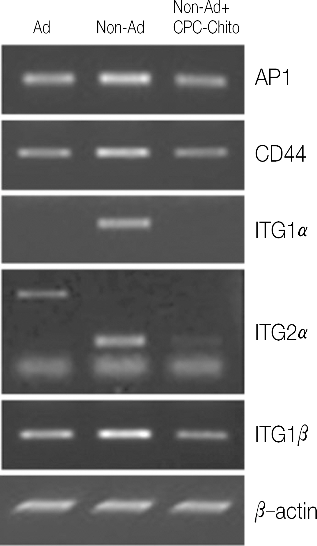
Fig. 5. The expression of mRNAs in MDPC-23 cells. Byung-Do Chun et al: Interaction between odontoblast and bio-calcium phosphate cement reinforced with chitosan. J Korean Assoc Oral Maxillofac Surg 2011
Reference
-
References
1. Pereira JC, Segala AD, Costa CAS. Human pulpal response to direct pulp capping with an adhesive system. Am J Dent. 2000; 13:139–47.2. Cox CF, Hafez AA, Akimoto N, et al. Biocompatibility of primer, adhesive and resin composite systems on non-exposed and exposed pulps of nonhuman primate teeth. Am J Dent. 1998; 11:S55–63.3. Machida Y, Nagai T, Abe M, Sannan T. Use of chitosan and hy-droxypropylchitosan in drug formulations to effect sustained release. Drug Dis Deliv. 1986; 1:119–30.4. Muzzarelli RAA, Biagini G, Bellardini M, Simonelli L, Castaldini C, Fratto G. Osteoconduction exerted by methylpyrro-lidinone chitosan used in dental surgery. Biomaterials. 1993; 14(1):39–43.
Article5. Muzzarelli RAA. Amphoteric derivatives of chitosan and their biological significance. Skjak-Brak G, Anthonsen T, Sandford P, editors. Chitin and chitosan. New York: Elsevier;1989. p. 87–99.6. Yamaguchi I, Tokuchi K, Fukuzaki H, Koyama Y, Takakuda K, Monma H, Tanaka J. Precipitation and microstructure analysis of chitosan/hydroxyapatite nanocomposites. J Biomed Mater Res. 2001; 55:20–7.7. LeGeros RZ. Biodegradation and bioresorption of calcium phosphate ceramics. Clin Mater. 1993; 14:65–88.
Article8. Chang MC, Ko CC, Douglas WH. Preparation of hydroxyapatite-gelatin nanocomposites. Biomaterials. 2003; 24:2853–62.9. Brown WE, Chow LC. A new calcium phosphate water setting cement. Brown PW, editor. Cements research progress. Westerville, OH: Am. Ceram. Soc.;1986. p. 352–79.10. Shindo ML, Contantino PD, Friedman CD, Chow LC. Facial skeletal augmentation using hydroxyapatite cement. Arch Otolaryngol Head Neck Surg. 1993; 119:185–90.
Article11. Friedman CD, Costantino PD, Takagi S, Chow LC. BoneSourceTM hydroxyapatite cement: a novel biomaterial for craniofacial skeletal tissue engineering and reconstruction. J Biomed Mater Res. 1998; 43B:428–32.12. Sugawara A, Kusama K, Nishimura S, Nishiyama M, Moro I, Kudo I, et al. Histopathological reaction of a calcium phosphate cement root canal filler. J Hard Tissue Biol. 1995; 4:1–7.13. Cherng AM, Chow LC, Takagi S. In vitro evaluation of a calcium phosphate cement root canal filler/sealer. J Endodontics. 2001; 27:613–5.14. Dickens-Venz SH, Takagi S, Chow LC, Bowen RL, Johnston AD, Dickens B. Physical and chemical properties of resin-reinforced calcium phosphate cements. Dent Mater. 1994; 10:100–6.
Article15. Dickens SH, Flaim GM, Takagi S. Mechanical properties and biochemical activity of remineralizing resin-based a-PO4 cements. Dent Mater. 2003; 19:558–66.16. Takagi S, Chow LC, Hirayama S, Eichmiller FC. Properties of novel resorbable chitosan-calcium phosphate composites. Dent Mater. 2003; 19:797–804.17. Xu HHK, Quinn JB, Takagi S, Chow LC. Synergistic reinforcement of in situ hardening calcium phosphate composite scaffold for bone tissue engineering. Biomaterials. 2004; 25:1029–37.
Article18. Xu HHK, Simon Jr CG. Fast setting calcium phosphate-chitosan scaffold: mechanical properties and biocompatibility. Biomaterials. 2005; 26:1337–48.
Article19. Di Martino A, Sittinger M, Risbud MV. Chitosan: a versatile biopolymer for orthopaedic tissue-engineering. Biomaterials. 2005; 26:5983–90.
Article20. Prabaharan M. Review paper: chitosan derivatives as promising materials for controlled drug delivery. J Biomater Appl. 2008; 23:5–36.
Article21. Kim IY, Seo SJ, Moon HS, Yoo MK, Park IY, Kim BC, et al. Chitosan and its derivatives for tissue engineering applications. Biotechnol Adv. 2008; 26:1–21.
Article22. Li X, Feng Q, Liu X, Dong W, Cui F. Collagen-based implants reinforced by chitin fibres in a goat shank bone defect model. Biomaterials. 2006; 27:1917–23.
Article23. Padois K, Rodriguez F. Effects of chitosan addition to self-setting bone cement. Biomed Mater Eng. 2007; 17:309–20.24. Khashaba RM, Chutkan NB, Borke JL. Comparative study of biocompatibility of newly developed calcium phosphate-based root canal sealers on fibroblasts derived from primary human gingiva and a mouse L929 cell line. Int Endod J. 2009; 42:711–8.
Article25. Dagang G, Kewei X, Haoliang S, et al. Physicochemical properties of TTCP/DCPA system cement formed in physiological saline solution and its cytotoxicity. J Biomed Mater Res A. 2006; 77:313–23.
Article26. Matsuya S, Takagi S, Chow LC. Effect of mixing ratio and pH on the reaction between Ca4 (PO4)2O and CaHPO4. J Mater Sci Mater Med. 2000; 11:305–11.27. Sun-Kyung Lee, Sang-Kwang Lee, Sang-Im Lee, Jeong-Hui Park, Jun-Hyeog Jang, Hae-Won Kim, Eun-Cheol Kim. Effect of Calcium Phosphate Cements on Growth and Odontoblastic Differentiation in Human Dental Pulp Cell. Journal of Endodontics. 2010; 36(9):1537–42.
- Full Text Links
-
- Actions
-
Cited
- CITED
-
- Close
- Share
-
- Similar articles
-
- Response of Odontoblast to the Bio-Calcium Phosphate Cement
- The experimental study of the effect of zinc phosphate cement on the solubility of enamel
- Surface characterization of calcium phosphate coating formed on chitosan and alkali-treaDted titanium metal
- Effects of Chitosan on the Differentiation of MDPC-23 Cells
- Two Cases of Cement Burn
