J Korean Assoc Oral Maxillofac Surg.
2010 Oct;36(5):438-440.
Lipoma on superficial lobe of the parotid gland: case report
- Affiliations
-
- 1Department of Oral and Maxillofacial Surgery, Department of Dentistry, Dong-A University Medical Center, Busan, Korea. samehope@naver.com
- 2Department of Oral and Maxillofacial Surgery, School of Dentistry, Pusan National University, Yangsan, Korea.
Abstract
- A lipoma is a benign tumor of matured adipose tissue that usually occurs at the shoulder, back, and abdomen. 13% of lipomas occur in the head and neck area. However, the incidence of lipoma in the parotid gland is very low, approximately 2.5%. A conservational surgical excision is recommended in cases of lipoma of the parotid gland, with only 1-2% of lipomas recurring. We report a case of a lipoma in the parotid gland that was removed by conservational surgical excision. The lesion was exposed by the pre-auricular approach and the tissue was detached. After the parotid gland envelop was exposed, a yellowish mass is observed that was easy to remove due to capsulation. Most authors recommend a surgical excision of the superficial lobe of the parotid gland as the treatment for a lipoma in the parotid gland. However, enucleation only may be a sufficient treatment when a lipoma occurs in the superficial lobe or around the parotid gland. A patidectomy is not needed when a lipoma is located at the superficial lobe of the parotid gland, and a conservational surgical excision is suitable. Therefore, a clinical diagnosis is important for reducing the damage to the facial nerve.
Keyword
Figure
-
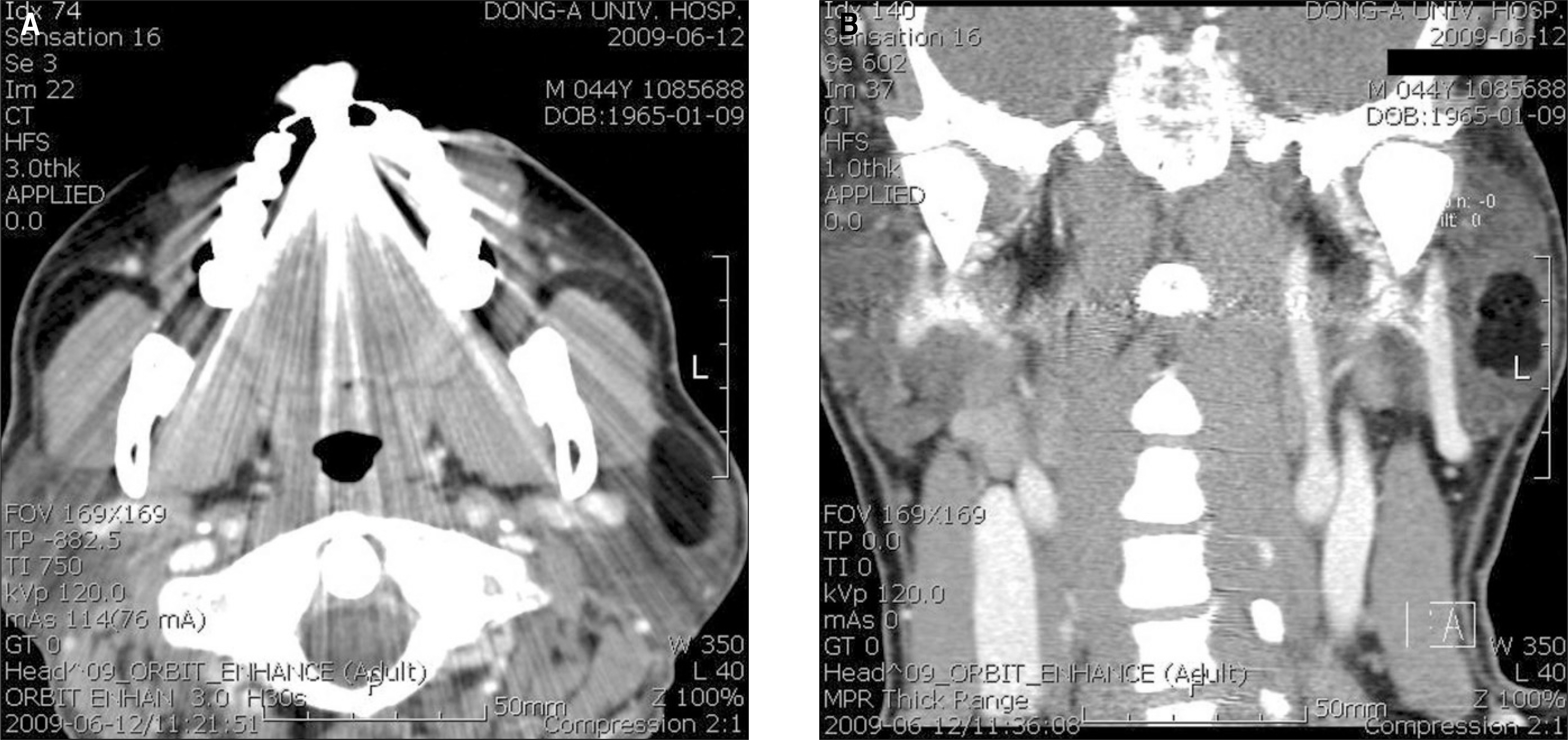
Fig. 1. Computed tomograph (CT) scan.(enhanced view) A. Transverse view of CT scan, B. Coronal view of CT scan.
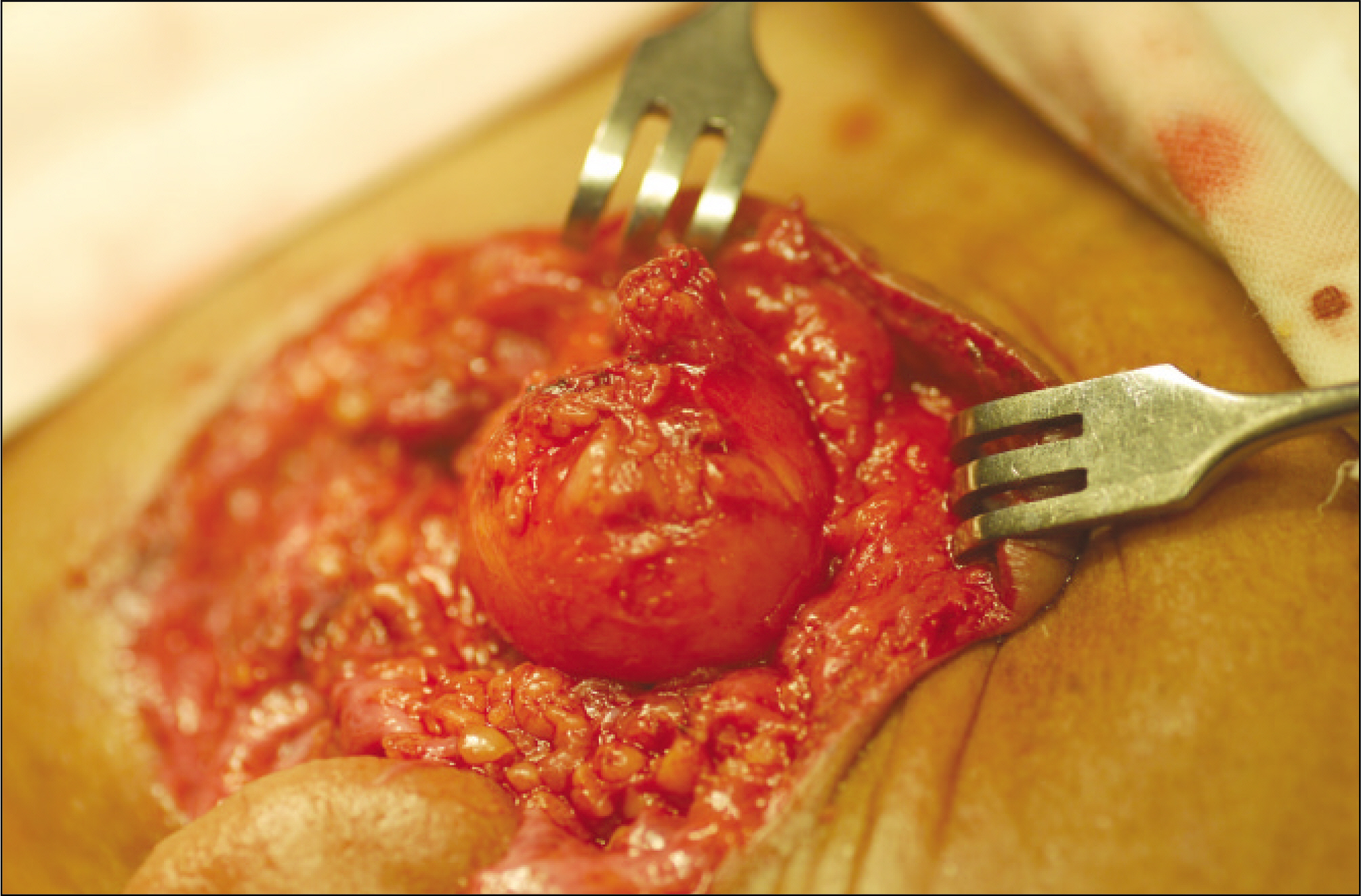
Fig. 2. Lipoma was exposed via preauricular approach.

Fig. 3. Macroscopic view (external) shows well-circumscribed, thinly encapsulated, and oval mass with soft consistency.

Fig. 4. Macroscopic view (cross section) shows uniform greasy cut surface with pale yellow color.
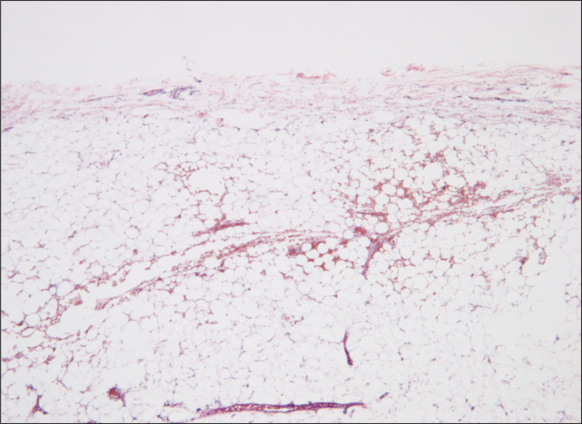
Fig. 5. The mass is well encapsulated by thin fibromembranous tissue.(H&E staining, original magnification ×40)
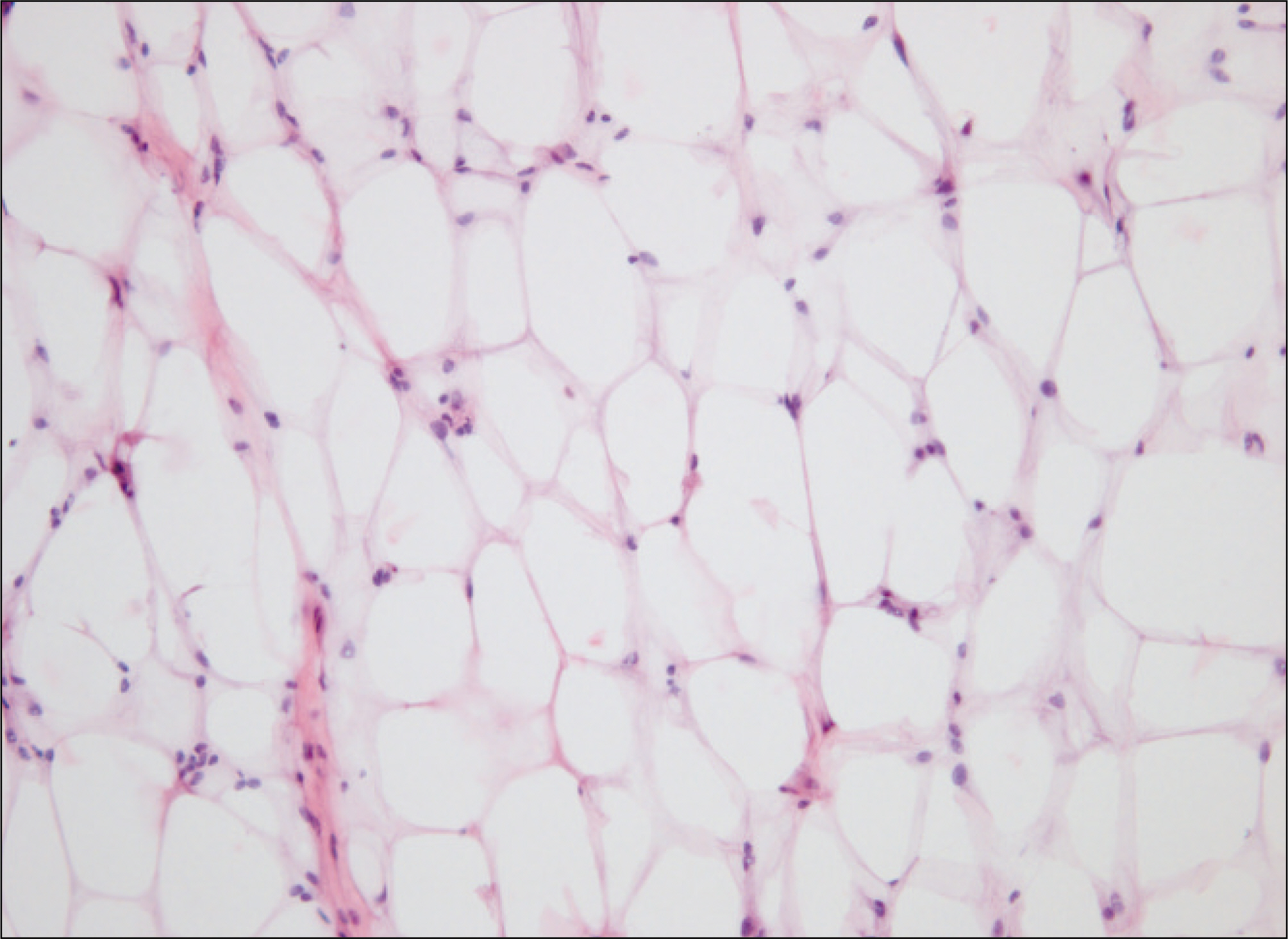
Fig. 6. The mass is composed of mature fat cells having only a slight variation in cellular size and shape.(H&E staining, original magnification ×200)
Reference
-
References
1. Williams TP, Stewart JC. Surgical pathology. Fonseca RJ, editor. Oral and Maxillofacial surgery. 1st ed.Philadelphia: W.B. Saunders;2000. p. 137–8.2. Som PM, Scherl MP, Rao VM, Biller HF. Rare presentations of ordinary lipomas of the head and neck: a review. AJNR Am J Neuroradiol. 1986; 7:657–64.3. Malave DA, Ziccardi VB, Greco R, Patterson GT. Lipoma of the parotid gland: report of a case. J Oral Maxillofac Surg. 1994; 52:408–11.
Article4. Walts AE, Perzik SL. Lipomatous lesions of the parotid area. Arch Otolaryngol. 1976; 102:230–2.
Article5. Kim YH, Reiner L. Ultrastructure of lipoma. Cancer. 1982; 50:102–6.
Article6. Houston GD, Brannon RB. Lipoma of the parotid gland. Oral Surg Oral Med Oral Pathol. 1985; 60:72–4.
Article7. Grage TB, Lober PH, Shahon DB. Benign tumors of the major salivary glands. Surgery. 1961; 50:625–33.8. Korentager R, Noyek AM, Chapnik JS, Steinhardt M, Luk SC, Cooter N. Lipoma and liposarcoma of the parotid gland: high resolution preoperative imaging diagnosis. Laryngoscope. 1988; 98:967–71.9. Sapp JP, Eversole LR, Wysocki GP. Contemporary oral and maxillofacial pathology. 2nd ed.St. Louis, Mo: Mosby;2004.10. Peel RL, Gnepp DR. Diseases of the salivary glands. Barnes L, editor. Surgical pathology of the head and neck. Vol. 1. 1st ed.New York, NY: Marcel Dekker;1985. p. 533–645.11. Janecka IP, Conley J, Perzin KH, Pitman G. Lipoma presenting as parotid tumors. Laryngoscope. 1977; 87:1007–10.12. Enzinger FM, Weiss SW. Soft tissue tumors. 2nd ed.St. Louis, Mo: Mosby;1988.
- Full Text Links
-
- Actions
-
Cited
- CITED
-
- Close
- Share
-
- Similar articles
-
- A Case of Lipoma of the Parotid Gland
- Lipoma of the parotid gland
- En-bloc Dissection of Deep and Superficial lobe of Parotid gland with Preserving the Facial Nerve
- A Case of Intraductal Papilloma arising in the Parotid Gland
- Epithelial-myoepithelial carcinoma on the superficial lobe of the parotid gland: a case report
