J Menopausal Med.
2014 Apr;20(1):39-42.
A Case of Ovarian Steroid Cell Tumor, Not Otherwise Specified, Treated with Surgery and Gonadotropin Releasing Hormone Agonist
- Affiliations
-
- 1Department of Pathology, Gachon University Gil Medical Center, Incheon, Korea.
- 2Department of Obstetrics and Gynecology, Gachon University Gil Medical Center, Incheon, Korea. miracle627@gilhospital.com
Abstract
- Steroid cell tumors account for less than 0.1% of all ovarian tumors. There are three steroid cell tumor subtypes: steroid cell tumor not otherwise specified (NOS), stromal luteoma and Leydig cell tumor. Steroid cell tumor, NOS, is the most common type and has malignant potential. This report describes a case of an ovarian steroid cell tumor, NOS. A 35-year-old woman visited hospital with the complaint of metrorrhagia. Physical examination revealed increased pubic hair. Transvaginal ultrasound indentified a 4.9 x 3.4 cm, well-circumscribed and solid left ovarian tumor. After laparoscopic left oophorectomy, the tumor was revealed as an ovarian steroid cell tumor, NOS. During the laparoscopic surgery, tumor ruptured. Complete surgical staging was performed and no evidence of metastasis was found. Gonadotropin releasing hormone agonist was administered monthly for 6 months. The patient has had no evidence of recurrence for 43 months.
MeSH Terms
Figure
-
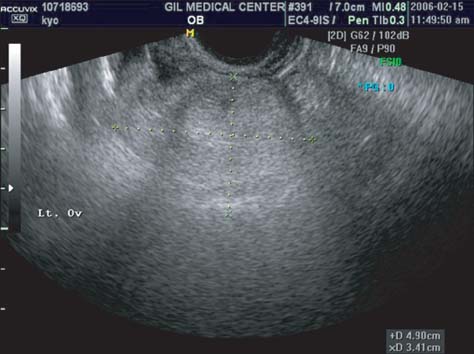
Fig. 1 Preoperative transvaginal ultrasonography. Four point nine times three point four cm sized solid tumor at left ovary.

Fig. 2 Microscopic photography of steroid cell tumor, not otherwise specified. (A) The tumor consists of irregular cords and nests of large rounded to polygonal cells (H&E, ×100). (B) The cytoplasm of the tumor cells show variable-sized clear vacuoles, representing fat material. Nuclei are monotonous, round and centrally located with prominent nucleoli (H&E, ×400).
Reference
-
1. Reedy MB, Richards WE, Ueland F, Uy K, Lee EY, Bryant C, et al. Ovarian steroid cell tumors, not otherwise specified: a case report and literature review. Gynecol Oncol. 1999; 75:293–297.2. Wang PH, Chao HT, Lee WL. Use of a long-acting gonadotropin-releasing hormone agonist for treatment of steroid cell tumors of the ovary. Fertil Steril. 1998; 69:353–355.3. Wang PH, Chao HT, Lee RC, Lai CR, Lee WL, Kwok CF, et al. Steroid cell tumors of the ovary: clinical, ultrasonic, and MRI diagnosis-a case report. Eur J Radiol. 1998; 26:269–273.4. Luk WT, Lee N, Chang TC, Chu KK. Lipid cell tumor of the ovary associated with endometrial adenocarcinoma-a case report. Changgeng Yi Xue Za Zhi. 1989; 12:244–248.5. Hayes MC, Scully RE. Ovarian steroid cell tumors (not otherwise specified). A clinicopathological analysis of 63 cases. Am J Surg Pathol. 1987; 11:835–845.6. Law KS, Chang TM, Tung JN. Fertility-sparing treatment of a primary retroperitoneal mucinous cystadenocarcinoma. BJOG. 2006; 113:612–614.7. Song MK, Lee YY, Lee KY, Lee A, Rha JG, Ryu KS, et al. A case of steroid cell tumor, not otherwise specified, with massive ascites. Korean J Obstet Gynecol. 2003; 46:2551–2555.8. Moon GS, Cho KH, Kim EJ, Chung JY, Nam ES, Cho SJ, et al. A case of lipid cell tumor of the ovary. Korean J Obstet Gynecol. 2004; 47:585–588.9. Oh JR, Cho SH, Yoon DK, Cho HB, Lee KE, Lee MB, et al. Laparoscopic management of asymptomatic ovarian steroid cell tumors, not otherwise specified: A case report and literature review. Korean J Obstet Gynecol. 2006; 49:933–938.10. Kim YT, Kim SW, Yoon BS, Kim SH, Kim JH, Kim JW, et al. An ovarian steroid cell tumor causing virilization and massive ascites. Yonsei Med J. 2007; 48:142–146.11. Lee JS, Kim BR, Lee HC, Lym BI, Kim HG, Moon HB. A case of ovarian steroid cell tumor, not otherwise specified with hypertension, obesity, ascites and elevated CA 125. Korean J Obstet Gynecol. 2008; 51:1164–1169.12. Park J, Kim TH, Lee HH, Lee W, Chung SH. Ovarian rete cyst in a post-menopausal woman: a case report. J Korean Soc Menopause. 2012; 18:67–69.13. Pascale MM, Pugeat M, Roberts M, Rousset H, Déchaud H, Dutrieux-Berger N, et al. Androgen suppressive effect of GnRH agonist in ovarian hyperthecosis and virilizing tumours. Clin Endocrinol (Oxf). 1994; 41:571–576.14. Imai A, Iida K, Tamaya T. Direct action of gonadotropinreleasing hormone (LH-RH) analogue on ovary: an alternative acting mechanism of buserelin. Arch Gynecol Obstet. 1991; 248:117–121.
- Full Text Links
-
- Actions
-
Cited
- CITED
-
- Close
- Share
-
- Similar articles
-
- A study of low dose purified follicle-stimulating hormone supplemented with gonadotropin releasing hormone agonist in women with polycystic ovarian disease
- Direct modulatory action of gonadotropin releasing hormone agonist on ovarian function : Effect on progesterone production in FSH-stumilated procine granulosa cell culture
- The effective of ultrashort-term gonadotropin-releasing hormone agonist administration in controlled ovarian hyperstimulation for in vitro fertilization
- Ovulation induction with combined gonadotropin-releasing hormone agonist(K-Trp-Luteinizing houmone-Releasing hormone:decapeptyl) and gonadotropins in women with polycystic ovarian disease
- Thyroid Dysfunction Associated with Administration of the Long-Acting Gonadotropin-Releasing Hormone Agonist
