J Korean Soc Spine Surg.
2004 Dec;11(4):246-252.
Epidemiologic study of lumbar scoliosis with plain abdominal X-ray
- Affiliations
-
- 1Seoul Spine Institute, Inje University Sanggye Paik Hospital, Korea. kjh1026@sanggyepaik.ac.kr
Abstract
- STUDY DESIGN: A retrospective cross-sectional study
OBJECTIVES
To analyze the prevalence and characteristics of lumbar scoliosis using plain abdominal X-rays, according to age. LITERATURE REVIEW SUMMARY: The single lumbar curves of adolescents have shown 10 ~20% idiopathic scoliosis, but the reported prevalence of adult lumbar scoliosis ranges from 2.5 to 7.5%. In Korea, there is no useful basic data concerning lumbar scoliosis.
MATERIALS AND METHODS
A total of 2877 plain abdominal radiographies (supine and erect), taken at our hospital, between August 2001 and June 2002, were retrospectively investigated. The ages of the patients ranged from 11 to 80 years, and the patients were grouped according to age. The prevalence, Cobb angle, ratio of males and females, ratio of right and left curves, location of end and apex vertebra, the number of involved vertebra in primary curve, amount of rotation and osteophytes were all examined.
RESULTS
The overall prevalence of lumbar scoliosis was 4.3% (N=124), but rapidly increased after the sixth decade. The average Cobb angle was 16.2 degree. A positive correlation was found between the Cobb angle and age (r=0.275, P<0.05). The ratios of males to females and of the right to left curves were both about 1:2. The most common sites of upper end vertebra were T12 and L1, that of the lower end vertebra L4 and those of the apex L2 (N=48) and L3 (N=40). Most (N=111) had grade 1 rotation. With regard to the magnitude of the curves, no other factors were statistically significant.
CONCLUSIONS
De novo scoliosis can be considered to develop rapidly after the sixth decade. The Cobb angle had a positive correlation with age (r=0.275, p<0.05). These data are thought could be useful and valuable for future study of lumbar scoliosis.
Keyword
MeSH Terms
Figure
-
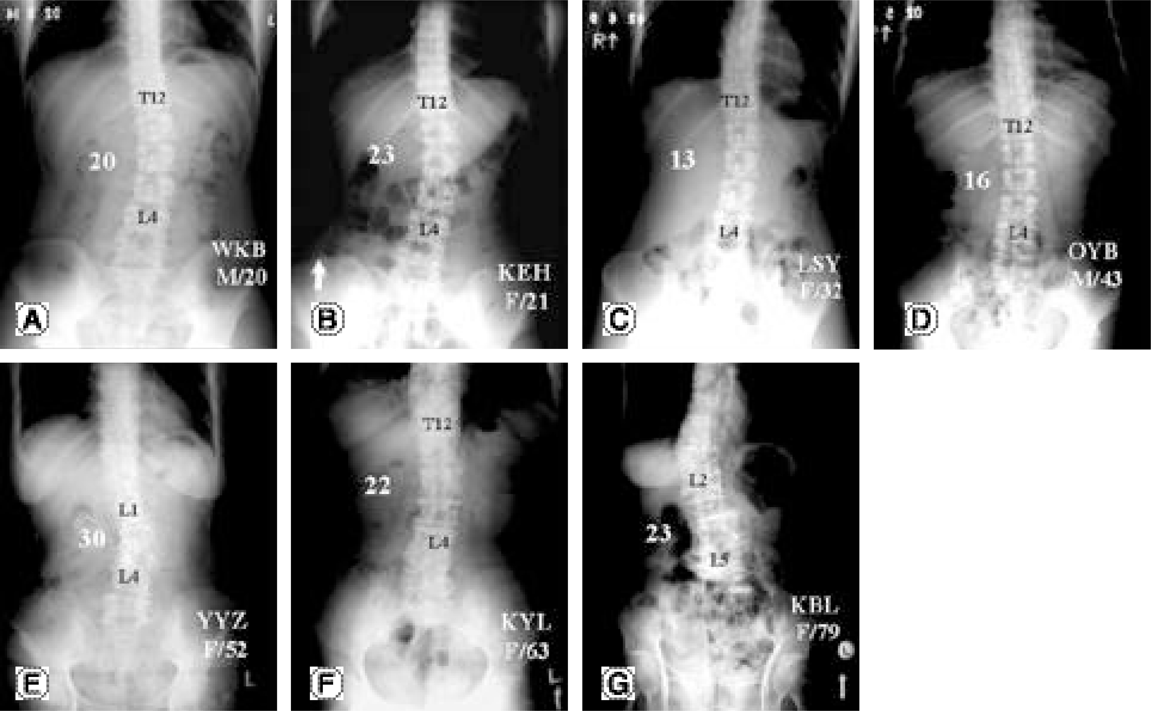
Fig. 1. These erect abdominal X-ray radiographies are examples of lumbar scoliosis grouped by age ((A) the second decade, (B) the third decade, (C) the fourth decade, (D) the fifth decade, (E) the sixth decade, (F) the seventh decade, (G) the eighth decade). The locations of apex are between the intervertebral disc space of L1 and L2 and body of L4. They show lumbar scoliosis of significant vertebral rotation and more than 10。

Fig. 2. The number of checked radiographies and lumbar scoliosis according to age.
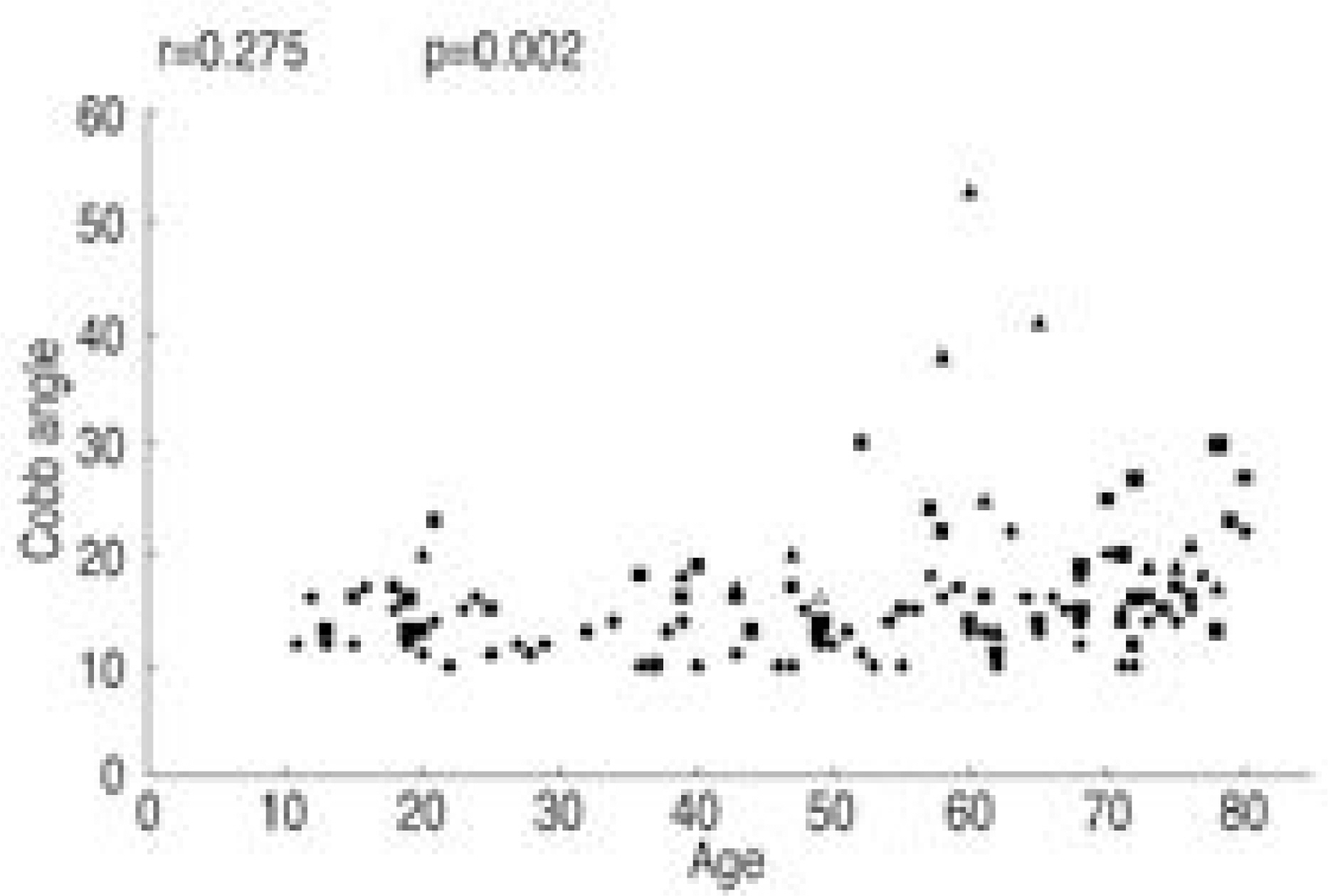
Fig. 3. Correlation between Cobb angle and age r: Pearson’s linear correlation coefficient
Reference
-
1). Kostuik JP, Bentivoglio J. The incidence of low-back pain in adult scoliosis. Spine. 1981; 8:268–273.
Article2). Cobb RJ. Outline for the study of scoliosis. Am Acad Orthop Surg. 194; 85:261–275.3). Nash CL, Moe J. A study of vertebral rotation. J Bone Joint Surg. 1969; 51-A:223–229.
Article4). Lonstein JE, Bjorklund S, Wanninger MH, Nelson RP. Voluntary school screening for scoliosis in Minnesota. J Bone Joint Surg. 1982; 64-A:481–488.
Article5). Renshaw TS. Screening school children for scoliosis. Clin Orthop. 1987; 229:26–33.
Article6). Rogala EJ, Drummond DS, Gurr J. Scoliosos: Incidence and natural history. A prospective epidemiological study. J Bone Joint Surg. 1978; 60-A:173–176.7). Suk SI, Choi IH, Ahn JW, Kim IK. The incidence of scoliosis in Korea part III: The incidence of scoliosis in the middle and high school students. J Kor Orthop Assoc. 1980; 15:1–6.
Article8). James JIP. Idiopathic scoliosis. The prognosis, diagnosis and operative indications related to curve patterns and the age at onset. J Bone Joint Surg. 1954; 36-B:36–49.9). Ponsei IV, Friedman B. Prognosis in idiopathic scoliosis. J Bone Joint Surg. 1950; 32-A:381–395.10). Cruickshank JL, Koike M, Dickson RA. Curve patterns in idiopathic scoliosis: A clinical and radiographic study. J Bone Joint Surg. 1989; 71-B:259–263.
Article11). Willner S, Uden A. A prospective prevalence study of scoliosis in southern Sweden. Acta Orthop Scand. 1982; 53:233–237.
Article12). Robin G, Span Y, Makin M, Steinberg R. Scoliosis in the elderly: Idiopathic or osteoporotic. Proceedings of the 5th Zorab scoliosis symposium. London. 1976.13). Perennou D, Marcelli C, Hrisson C, Simon L. Adult lumbar scoliosis: Epidemiologic aspects in a low-back pain population. Spine. 1994; 19:123–128.14). Pritchett JW, Bortel DT. Degenerative symptomatic lumbar scoliosis. Spine. 1993; 18:700–703.
Article15). Moon MS, Lee KS, Lim CI, Kim YB, Lee HS. A clinical study of degerative lumbar scoliosis. J Kor Orthop Assoc. 1992; 27:946–951.16). Korovessis P, Piperos G, Sidiropoulos P, Dimas A. Adult idiopathic lumbar scoliosis: A formula for prediction of progression and review of the literature. Spine. 1994; 19:1926–1932.17). Schwab FJ, Smith VA, Biserni M, Gamez L, Farcy JC, Pagala M. Adult Scoliosis: A quantitative radiographic and clinical analysis. Spine. 2002; 27:387–392.
- Full Text Links
-
- Actions
-
Cited
- CITED
-
- Close
- Share
-
- Similar articles
-
- Clinico-radiologic Findings of the Whole Spine in Patients with Chronic Low Back Pain
- The Usefulness of Chest and Abdominal CT in Detection of Thoracolumbar Spine Fracture after Multiple Blunt Trauma
- Plain Abdominal Radiography in Infants and Children
- Lumbar Scoliosis in Patients With Breast Cancer: Prevalence and Relationship With Breast Cancer Treatment, Age, Bone Mineral Density, and Body Mass Index
- Plain abdominal and chest findings of ruptured ectopic pregnancies
