Unusual Primary Subepithelial Tumors of the Colon: Multimodality Imaging Findings with Endoscopic and Pathologic Correlation
- Affiliations
-
- 1Department of Diagnostic Radiology, Dong-A University College of Medicine, Busan, Korea. medcarrot@naver.com
- 2Department of Internal Medicine, Dong-A University College of Medicine, Busan, Korea.
- 3Department of Diagnostic Radiology, Pusan National University College of Medicine, Busan, Korea.
Abstract
- The colonoscopy has been used to diagnose various colonic lesions. However, this method has its limitations in diagnosing and differentiating subepithelial tumors. For this reason, the role of cross-sectional radiologic imaging is important for the diagnosis of colonic subepithelial tumors. Moreover, although these tumors are associated with a wide range of radiologic features, they may have unique radiologic features that suggest a specific diagnosis. Hemangiomas typically show transmural colonic wall thickening with phleboliths in intramural or extracolic areas. Colonic lymphangiomas manifest as a multilocular cystic mass at CT and sonography. Colonic lipomas are well demonstrated by CT because the masses were present with characteristic fatty density. Schwannomas usually appear as well circumscribed, homogeneous masses with low attenuation at CT. The primary form of colonic lymphoma has a wide variety of radiologic types, including a polypoid mass, circumferential mural mass, and a cavitary mass. Small gastrointestinal stromal tumors are usually homogeneous, whereas larger tumors tend to have a heterogeneous appearance with central necrosis at contrast-enhanced CT scans. Neuroendocrine tumors of the colon are most frequently observed in the rectum and are typically small incidental lesions. Familiarity with these imaging features can help distinguish particular disease entities.
MeSH Terms
Figure
-
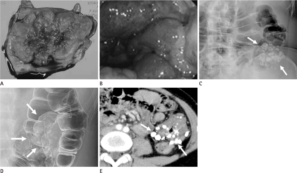
Fig. 1 Hemangioma of the descending colon in a 34-year-old woman. A. Photograph of the resected specimen demonstrates a lobulated subepithelial mass with areas of focal hemorrhage and multiple phleboliths. B. Colonoscopic image shows dilated subepithelial vascular masses with a deep wine color associated with mucosal congestion and edema. C. Plain radiograph shows multiple phleboliths (arrows) along the descending colon. D. Barium study reveals lobulated contour masses (arrows) with multiple phleboliths along the proximal portion of the descending colon. E. Contrast-enhanced CT scan reveals circumferential mural thickening with multiple phleboliths in the descending colon (arrows).
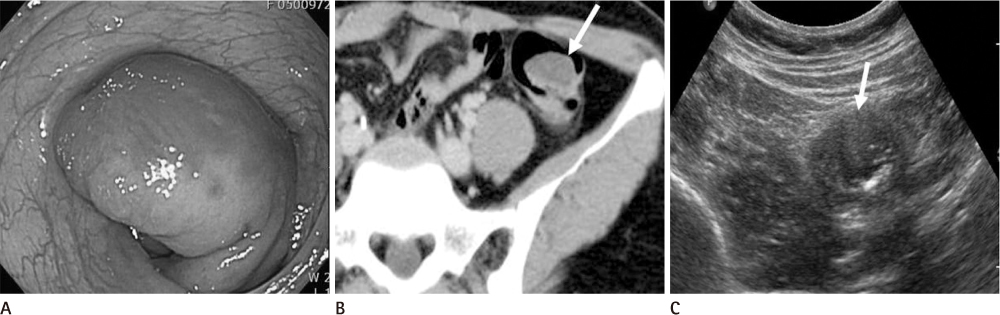
Fig. 2 Lymphangioma of the colon in a 33-year-old woman. A. Photography from endoscopy reveals a pedunculated subepithelial mass covered with erythematous mucosa in the sigmoid colon. B. Contrast-enhanced CT scan shows a well-defined low attenuated cystic mass in the sigmoid colon (arrow). C. Transabdominal sonogram shows an anechoic cystic mass in the sigmoid colon (arrow).
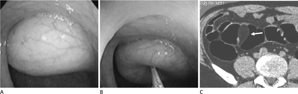
Fig. 3 Lipoma of the sigmoid colon in a 51-year-old man. A. Colonoscopic image shows a yellowish subepithelial mass. B. The mass reveals a cushion sign at compression with a blunt instrument. C. Axial CT scan shows a well-defined fatty mass in the sigmoid colon (arrow).
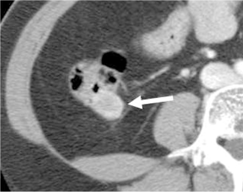
Fig. 4 Schwannoma of the cecum in a 44-year-old man. Contrast-enhanced CT scan shows a homogeneously enhancing, exoenteric growing mass (arrow) in the posterior wall of the cecum.

Fig. 5 Inflammatory fibroid polyp of the ascending colon in a 35-year-old man. A. Photograph from endoscopy reveals a subepithelial mass with a smooth mucosal surface (arrow). B. Contrast-enhanced CT shows a well-defined, homogeneously low attenuated mass with intact overlying mucosa (arrow) in the ascending colon.

Fig. 6 Polypoid primary lymphoma of the cecum in an 18-year-old man. A. Barium study reveals a lobulated filling defect with a "coiled spring" appearance in the hepatic flexure of the colon (arrow). B. Transabdominal sonogram shows a well-defined hypoechoic mass (arrow) with intussusception of the ascending colon. C. Corresponding contrast-enhanced CT scan shows the inhomogeneously enhancing mass combined with intussusception within the ascending colon. D. Photograph of the resected specimen shows a well-demarcated fungating mass with focal ulceration in the cecum.
Fig. 7 Circumferential infiltrative primary lymphoma of the rectum in a 29-year-old man. A. Pre-contrast CT scan shows low attenuated circumferential mural thickening of the rectum. B. Contrast-enhanced CT scan reveals poor contrast enhancement of the lesion. C. Barium study demonstrates segmental luminal narrowing with lobulated contour defect (arrows) in the rectum.

Fig. 8 Gastrointestinal stromal tumor of the rectum in a 56-year-old man. A. Photograph of the resected specimen shows intramural mass with intact overlying mucosa in the rectal wall. B. Contrast-enhanced CT scan shows an exoenteric growing mass with inhomogeneous enhancement in the anterior wall of the rectum. C. T1-weighted axial image. D. T2-weighted sagittal image. E. Gd-enhanced fat suppressed T1-weighted image demonstrate a well-defined exoenteric subepithelial mass with heterogeneous enhancement (arrows).
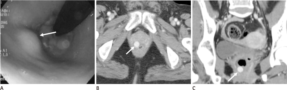
Fig. 9 Neuroendocrine tumor of the rectum in a 55-year-old woman. A. Colonoscopic image reveals a small subepithelial mass (arrow) with intact overlying mucosa in the lower rectum. Axial (B) and coronal (C) reformatted CT images show a small well-enhancing subepithelial mass (arrows) in the lower rectum.
Fig. 10 Leiomyosarcoma of the rectum in a 64-year-old man. On contrast-enhanced CT scan, a well-circumscribed, moderately enhancing mural mass with focal necrosis is noted in the rectum.
Reference
-
1. Pickhardt PJ, Kim DH, Menias CO, Gopal DV, Arluk GM, Heise CP. Evaluation of submucosal lesions of the large intestine: part 1. Neoplasms. Radiographics. 2007; 27:1681–1692.2. Hsu RM, Horton KM, Fishman EK. Diffuse cavernous hemangiomatosis of the colon: findings on three-dimensional CT colonography. AJR Am J Roentgenol. 2002; 179:1042–1044.3. Wan YL, Lee TY, Hung CF, Ng KK. Ultrasound and CT findings of a cecal lymphangioma presenting with intussusception. Eur J Radiol. 1998; 27:77–79.4. Genchellac H, Demir MK, Ozdemir H, Unlu E, Temizoz O. Computed tomographic and magnetic resonance imaging findings of asymptomatic intra-abdominal gastrointestinal system lipomas. J Comput Assist Tomogr. 2008; 32:841–847.5. Levy AD, Quiles AM, Miettinen M, Sobin LH. Gastrointestinal schwannomas: CT features with clinicopathologic correlation. AJR Am J Roentgenol. 2005; 184:797–802.6. Harned RK, Buck JL, Shekitka KM. Inflammatory fibroid polyps of the gastrointestinal tract: radiologic evaluation. Radiology. 1992; 182:863–866.7. Ghai S, Pattison J, Ghai S, O'Malley ME, Khalili K, Stephens M. Primary gastrointestinal lymphoma: spectrum of imaging findings with pathologic correlation. Radiographics. 2007; 27:1371–1388.8. Levy AD, Remotti HE, Thompson WM, Sobin LH, Miettinen M. radiologic features with pathologic correlation. Radiographics. 2003; 23:283–304. 456quiz 532.9. Levy AD, Remotti HE, Thompson WM, Sobin LH, Miettinen M. Anorectal gastrointestinal stromal tumors: CT and MR imaging features with clinical and pathologic correlation. AJR Am J Roentgenol. 2003; 180:1607–1612.10. Levy AD, Sobin LH. From the archives of the AFIP: Gastrointestinal carcinoids: imaging features with clinicopathologic comparison. Radiographics. 2007; 27:237–257.
- Full Text Links
-
- Actions
-
Cited
- CITED
-
- Close
- Share
-
- Similar articles
-
- Endoscopic Full-Thickness Resection for Gastric Subepithelial Lesions Arising from the Muscularis Propria
- Contrast Enhanced Endoscopic Ultrasound Imaging for Gastrointestinal Subepithelial Tumors
- Endoscopic Treatment of Subepithelial Tumors
- Endoscopic Management of Gastric Subepithelial Tumor
- Splenic Cyst Mimicking Gastric Subepithelial Tumor Diagnosed by Endoscopic Ultrasonography
